
Page 346 - Equine Clinical Medicine, Surgery and Reproduction, 2nd Edition
P. 346
Musculoskeletal system: 1.8 Soft-tissue injuries 321
VetBooks.ir or metatarsal bones) are probably significant and Computed tomography
There have been very few reports of the use of CT
associated with a poorer prognosis for return to
full function and increased risk of recurrence
resolution, especially of mineralised tissues. It will
(Fig. 1.623). These may be difficult to visualise for this condition, although CT offers high spatial
ultrasonographically, although examining the limb show bone sclerosis deep within the proximal pal-
non-weight bearing while passively moving the mar third metacarpal/plantar third metatarsal bones
lower leg may help to visualise fibrous, restrictive with increased bone density, loss of trabecular pat-
adhesions. tern and thickening of the cortex. Entheseophytes
are clearly visible. Fissure lines or avulsion frag-
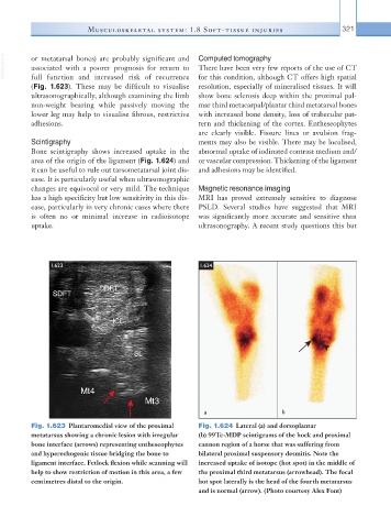
Scintigraphy ments may also be visible. There may be localised,
Bone scintigraphy shows increased uptake in the abnormal uptake of iodinated contrast medium and/
area of the origin of the ligament (Fig. 1.624) and or vascular compression. Thickening of the ligament
it can be useful to rule out tarsometatarsal joint dis- and adhesions may be identified.
ease. It is particularly useful when ultrasonographic
changes are equivocal or very mild. The technique Magnetic resonance imaging
has a high specificity but low sensitivity in this dis- MRI has proved extremely sensitive to diagnose
ease, particularly in very chronic cases where there PSLD. Several studies have suggested that MRI
is often no or minimal increase in radioisotope was significantly more accurate and sensitive than
uptake. ultrasonography. A recent study questions this but
1.623 1.624
a b
Fig. 1.623 Plantaromedial view of the proximal Fig. 1.624 Lateral (a) and dorsoplantar
metatarsus showing a chronic lesion with irregular (b) 99Tc-MDP scintigrams of the hock and proximal
bone interface (arrows) representing entheseophytes cannon region of a horse that was suffering from
and hyperechogenic tissue bridging the bone to bilateral proximal suspensory desmitis. Note the
ligament interface. Fetlock flexion while scanning will increased uptake of isotope (hot spot) in the middle of
help to show restriction of motion in this area, a few the proximal third metatarsus (arrowhead). The focal
centimetres distal to the origin. hot spot laterally is the head of the fourth metatarsus
and is normal (arrow). (Photo courtesy Alex Font)