
Page 369 - Equine Clinical Medicine, Surgery and Reproduction, 2nd Edition
P. 369
344 CHAPTER 1
VetBooks.ir (‘sympathetic’) effusion within the carpal flexor ten- be effective. Corrective farriery should include
keeping the toe short with rolling of the toe of
don sheath. With time, the echogenicity increases
but the ligament remains markedly thickened
Heel wedges have been suggested but should be
(Fig. 1.677). Adhesions with the deep and superficial the shoe. The heels should be kept slightly long.
digital flexor tendons are frequent, especially laterally avoided as they may cause retraction of the liga-
and in chronic cases (Figs. 1.676, 1.677). ment and flexural deformity. If the latter occurs,
desmotomy of the check ligament and use of toe-
Management extension shoes are indicated. Use of intralesional
Rest and conservative treatment as for tendon- injections of biological products (PRP, stem cells,
itis are usually effective, and the lesions tend to UBM) are probably not indicated but have been
heal more rapidly than in the SDFT. Aggressive advocated by some authors.
physiotherapy with controlled exercise and pas-
sive manipulations aimed at decreasing restric- Prognosis
tive adhesion formation are useful. In chronic, Recurrence is common and the desmitis then tends
recurrent cases, surgical resection (desmotomy) to evolve into a chronic, recurrent form. The prog-
of a 5 cm long portion of the check ligament may nosis is fair in acute, mild cases, but more guarded in
1.676 1.677
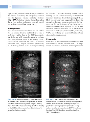
Fig. 1.676 Severe diffuse injury in the distal part Fig. 1.677 A chronic/healed injury where the
of the AL-DDFT with near complete loss of normal echogenicity is near normal, although heterogeneous,
structure and loss of the ligament margins (arrows). and the ligament remains markedly enlarged with
Note the mottled tissue extending laterally to the edge poorly defined margins (arrows). Note the poorly
of the SDFT, with associated loss of definition of its defined interface between the AL-DDFT, the dorsal
lateral border (arrowhead). border of the DDFT and the lateral border of the
SDFT (arrowheads).