
Page 373 - Equine Clinical Medicine, Surgery and Reproduction, 2nd Edition
P. 373
348 CHAPTER 1
VetBooks.ir 1.685
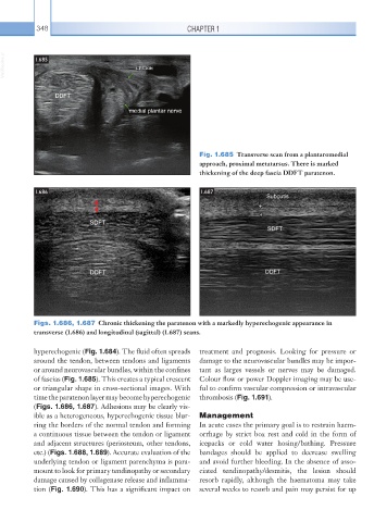
Fig. 1.685 Transverse scan from a plantaromedial
approach, proximal metatarsus. There is marked
thickening of the deep fascia DDFT paratenon.
1.686 1.687
Figs. 1.686, 1.687 Chronic thickening the paratenon with a markedly hyperechogenic appearance in
transverse (1.686) and longitudinal (sagittal) (1.687) scans.
hyperechogenic (Fig. 1.684). The fluid often spreads treatment and prognosis. Looking for pressure or
around the tendon, between tendons and ligaments damage to the neurovascular bundles may be impor-
or around neurovascular bundles, within the confines tant as larges vessels or nerves may be damaged.
of fascias (Fig. 1.685). This creates a typical crescent Colour flow or power Doppler imaging may be use-
or triangular shape in cross-sectional images. With ful to confirm vascular compression or intravascular
time the paratenon layer may become hyperechogenic thrombosis (Fig. 1.691).
(Figs. 1.686, 1.687). Adhesions may be clearly vis-
ible as a heterogeneous, hyperechogenic tissue blur- Management
ring the borders of the normal tendon and forming In acute cases the primary goal is to restrain haem-
a continuous tissue between the tendon or ligament orrhage by strict box rest and cold in the form of
and adjacent structures (periosteum, other tendons, icepacks or cold water hosing/bathing. Pressure
etc.) (Figs. 1.688, 1.689). Accurate evaluation of the bandages should be applied to decrease swelling
underlying tendon or ligament parenchyma is para- and avoid further bleeding. In the absence of asso-
mount to look for primary tendinopathy or secondary ciated tendinopathy/desmitis, the lesion should
damage caused by collagenase release and inflamma- resorb rapidly, although the haematoma may take
tion (Fig. 1.690). This has a significant impact on several weeks to resorb and pain may persist for up