
Page 374 - Equine Clinical Medicine, Surgery and Reproduction, 2nd Edition
P. 374
Musculoskeletal system: 1.8 Soft-tissue injuries 349
VetBooks.ir 1.688 1.689
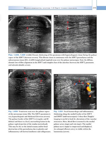
Figs. 1.688, 1.689 (1.688) Chronic thickening of the paratenon with hyperechogenic tissue lining the palmar
aspect of the SDFT (between arrows). This fibrous tissue is continuous with the SDFT parenchyma and the
subcutaneous tissue (SC). (1.689) Longitudinal (sagittal) scan over the palmar metacarpus. Note the diffuse,
chronic loss of fibre alignment in the SDFT and complete loss of the interface between the SDFT, paratenon
and subcutis (double arrow).
1.690
1.691
Fig. 1.690 Transverse scan over the palmar aspect Fig. 1.691 Focal haemorrhage and inflammatory
of the metacarpus (zone IIb). The SDFT paratenon is thickening along the medial border of the SDFT
very hypoechogenic and thickened (between arrows). and DDFT (mid-metacarpus). Colour-flow Doppler
The palmar border of the SDFT is irregular and ill imaging is useful to look for alterations of the vascular
defined and there is a focal, encroaching lesion in the structures. Here, blood flow is normal in the medial
palmar sagittal portion of the tendon (arrowhead). palmar artery (mpa) and vein (mpv). The medial
This may be due to the initial trauma and/or focal palmar nerve (mpn) is compressed and displaced.
destruction of the parenchyma due to platelet and An enlarged efferent artery is visible within the
inflammatory cell derived mediators and collagenases. haematoma (arrow).