
Page 639 - Equine Clinical Medicine, Surgery and Reproduction, 2nd Edition
P. 639
614 CHAPTER 3
VetBooks.ir 3.38 Radiography may be used, including lesion specific
obliques, but it is often difficult to interpret the results.
It is surprising how subtle some facial fractures are
on radiographs, particularly due to the complicated
anatomy and the thin nature of the bone. CT scans
are much more reliable, if available. It is important to
assess the paranasal sinuses for the presence of blood,
often seen as fluid lines in one or more of the sinuses.
Damage to the orbit or cranial vault should also be
considered. Ultrasonography of fractures and swell-
ings can be used, although it may be complicated by
the hyperechoic artefact from subcutaneous gas.
Management
Small skin punctures should be cleaned regularly
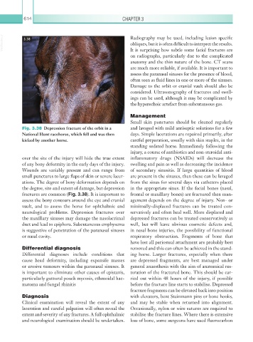
Fig. 3.38 Depression fracture of the orbit in a and lavaged with mild antiseptic solutions for a few
National Hunt racehorse, which fell and was then days. Simple lacerations are repaired primarily, after
kicked by another horse. careful preparation, usually with skin staples, in the
standing sedated horse. Immediately following the
injury, a course of antibiotics and non-steroidal anti-
over the site of the injury will hide the true extent inflammatory drugs (NSAIDs) will decrease the
of any bony deformity in the early days of the injury. swelling and pain as well as decreasing the incidence
Wounds are variably present and can range from of secondary sinusitis. If large quantities of blood
small punctures to large flaps of skin or severe lacer- are present in the sinuses, then these can be lavaged
ations. The degree of bony deformation depends on from the sinus for several days via catheters placed
the degree, site and extent of damage, but depression in the appropriate sinus. If the facial bones (nasal,
fractures are common (Fig. 3.38). It is important to frontal or maxillary bones) are fractured then man-
assess the bony contours around the eye and cranial agement depends on the degree of injury. Non- or
vault, and to assess the horse for ophthalmic and minimally-displaced fractures can be treated con-
neurological problems. Depression fractures over servatively and often heal well. More displaced and
the maxillary sinuses may damage the nasolacrimal depressed fractures can be treated conservatively as
duct and lead to epiphora. Subcutaneous emphysema well, but will leave obvious cosmetic defects and,
is suggestive of penetration of the paranasal sinuses in nasal bone injuries, the possibility of functional
or nasal cavity. respiratory obstruction. Fragments of bone that
have lost all periosteal attachment are probably best
Differential diagnosis removed and this can often be achieved in the stand-
Differential diagnoses include conditions that ing horse. Larger fractures, especially when there
cause head deformity, including expansile masses are depressed fragments, are best managed under
or erosive tumours within the paranasal sinuses. It general anaesthesia with the aim of anatomical res-
is important to eliminate other causes of epistaxis, toration of the fractured bone. This should be car-
particularly guttural pouch mycosis, ethmoidal hae- ried out within 48 hours of the injury, if possible
matoma and fungal rhinitis before the fracture line starts to stabilise. Depressed
fracture fragments can be elevated back into position
Diagnosis with elevators, bent Steinmann pins or bone hooks,
Clinical examination will reveal the extent of any and may be stable when returned into alignment.
laceration and careful palpation will often reveal the Occasionally, nylon or wire sutures are required to
extent and severity of any fractures. A full ophthalmic stabilise the fracture lines. Where there is extensive
and neurological examination should be undertaken. loss of bone, some surgeons have used fluorocarbon