
Page 641 - Equine Clinical Medicine, Surgery and Reproduction, 2nd Edition
P. 641
616 CHAPTER 3
VetBooks.ir Diagnosis Clinical presentation
Horses affected are usually mature, but otherwise no
Clinical examination of the nostrils may reveal
thickened and raised nodular masses in the mucosa,
tified. The clinical presentation is of a low-grade,
often with areas of haemorrhage. Endoscopy can breed, age or occupation risk factors have been iden-
be used to examine the rest of the URT for their recurrent, unilateral epistaxis or a serosanguineous,
presence elsewhere. Biopsy and histopathology will non-odourous nasal discharge (Fig. 3.41). The blood
confirm the diagnosis. Any generalised signs should in the discharge is old and not related to exercise.
be investigated thoroughly before treating the nasal Occasional cases present with airway obstruction,
lesions. abnormal respiratory sounds at exercise and poor
performance. Larger masses can cause facial defor-
Management mity, spread into the pharynx causing dysphagia, or
The underlying primary disease needs to be identi- extend down the nasal passages to become visible at
fied and treated but this is not detectable in some the nares. Rare cases have been associated with neu-
cases. Local surgical excision of the nostril lesions rological signs due to expansion through the cribri-
can be undertaken, and laser ablation of the nasal and form plate and head shaking.
other URT lesions is possible transendoscopically.
Differential diagnosis
Prognosis The principal differential diagnosis for investigation
The local URT lesions can recur and carry a guarded is epistaxis, especially guttural pouch mycosis. The
prognosis, while the primary problem can lead to a clinical presentation is, however, quite different.
poor prognosis.
PROGRESSIVE ETHMOIDAL 3.41
HAEMATOMA (PEH)
Definition/overview
Ethmoidal haematoma develops from the nasal or
sinus surface of the ethmoidal labyrinth and behaves
as a tumour, being progressive and expansile, but
histology does not reveal any neoplastic tissue. Most
commonly affected horses present with a low-grade
nasal discharge, often with blood, and are diagnosed
by endoscopy and radiography. A variety of options
are available for their treatment.
Aetiology/pathophysiology
The aetiology is unknown, but it is not neoplas-
tic. The pathophysiology is of repeated submuco-
sal haemorrhage from the surface of the ethmoidal
labyrinth, underneath normal respiratory epithe-
lium, and associated with significant fibrosis. The
mass itself is a mixture of blood, haemosiderin and
fibrous tissue with macrophages and giant cells. It is
unknown why the haemorrhage is repetitive, result-
ing in progression of the lesion. The masses can
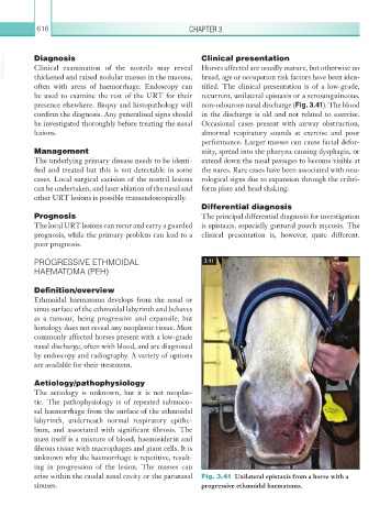
arise within the caudal nasal cavity or the paranasal Fig. 3.41 Unilateral epistaxis from a horse with a
sinuses. progressive ethmoidal haematoma.