
Page 642 - Equine Clinical Medicine, Surgery and Reproduction, 2nd Edition
P. 642
Respir atory system: 3.2 Surgical conditions of the respir atory tr act 617
VetBooks.ir More clinically typical differential diagnoses include endoscopy unless they invade through the conchae.
Ethmoidal haematomas visible on nasal endoscopy
trauma, mycotic rhinitis and sinonasal neoplasia.
Diagnosis are often associated with contralateral haematoma
within the paranasal sinuses. Bilateral sinusoscopy,
Endoscopic examination is diagnostic for lesions that via a maxillary or frontal trephine, is therefore indi-
arise from the nasal surface of the ethmoid (Fig. 3.42). cated in all cases of suspected or confirmed ethmoidal
Ethmoidal haematoma can be smooth-walled and haematoma (Fig. 3.43).
are usually reddish green/yellow in colour. Some Standing radiography of the head in a lateral
large lesions can enlarge around the caudal part of projection can be useful in the diagnosis of PEH,
the nasal septum and appear on endoscopy of the con- especially in sinus or larger lesions. The appearance
tralateral nasal passage. Bilateral lesions are frequent is of a soft-tissue mass, usually with quite a defined
and should be checked for in each case. Paranasal outline, rostral or dorsal to the ethmoidal labyrinth
sinus lesions are unlikely to be visible on per nasum (Fig. 3.44). Dorsoventral views may confirm its
3.42 3.43
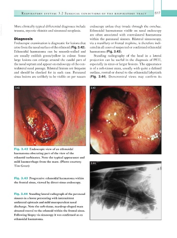
Fig. 3.42 Endoscopic view of an ethmoidal
haematoma obscuring part of the view of the
ethmoid turbinates. Note the typical appearance and
mild haemorrhage from the mass. (Photo courtesy
3.44
Tim Greet)
Fig. 3.43 Progressive ethmoidal haematoma within
the frontal sinus, viewed by direct sinus endoscopy.
Fig. 3.44 Standing lateral radiograph of the paranasal
sinuses in a horse presenting with intermittent
unilateral epistaxis and mild mucopurulent nasal
discharge. Note the soft-tissue, teardrop-shaped mass
situated rostral to the ethmoid within the frontal sinus.
Following biopsy via sinuscopy it was confirmed as an
ethmoidal haematoma.