
Page 651 - Equine Clinical Medicine, Surgery and Reproduction, 2nd Edition
P. 651
626 CHAPTER 3
VetBooks.ir 3.56 3.57
Fig. 3.56 A large subepiglottic cyst. Fig. 3.57 A subepiglottic cyst just visible above the
(dorsally displaced) soft palate.
3.58
a lateral radiograph of the oropharynx (especially
if taken with the mouth open) reveals a triangular-
shaped gas radiolucency. Occasionally, a rounded
soft-tissue density is observed, which is typical
of a subepiglottic cyst (Fig. 3.58). In many cases
there is no gas between the base of the tongue and
the epiglottis. In this case the radiograph is non-
diagnostic – many normal horses have no gas here
and the epiglottic cyst is the same density as the
surrounding tissues.
Endoscopy via the mouth is seldom required if
a careful pharyngeal examination is performed,
including manipulation under local anaesthesia.
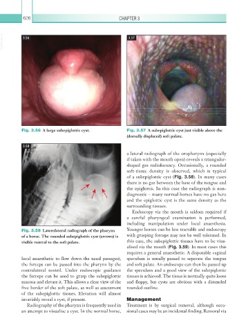
Fig. 3.58 Laterolateral radiograph of the pharynx Younger horses can be less tractable and endoscopy
of a horse. The rounded subepiglottic cyst (arrows) is with grasping forceps may not be well tolerated. In
visible ventral to the soft palate. this case, the subepiglottic tissues have to be visu-
alised via the mouth (Fig. 3.59). In most cases this
requires a general anaesthetic. A disposable vaginal
local anaesthetic to flow down the nasal passages), speculum is usually passed to separate the tongue
the forceps can be passed into the pharynx by the and soft palate. An endoscope can then be passed up
contralateral nostril. Under endoscopic guidance the speculum and a good view of the subepiglottic
the forceps can be used to grasp the subepiglottic tissues is achieved. The tissue is normally quite loose
mucosa and elevate it. This allows a clear view of the and floppy, but cysts are obvious with a distended
free border of the soft palate, as well as assessment rounded outline.
of the subepiglottic tissues. Elevation will almost
invariably reveal a cyst, if present. Management
Radiography of the pharynx is frequently used in Treatment is by surgical removal, although occa-
an attempt to visualise a cyst. In the normal horse, sional cases may be an incidental finding. Removal via