
Page 654 - Equine Clinical Medicine, Surgery and Reproduction, 2nd Edition
P. 654
Respir atory system: 3.2 Surgical conditions of the respir atory tr act 629
VetBooks.ir Clinical presentation 3.63
Nasal and nasopharyngeal foreign bodies can pres-
ent with marked epistaxis, particularly while the
horse is being exercised, due to damage to the nasal
turbinates. Coughing can be present, and horses can
be quite distressed with palpable throat pain and
resistance to flexion of the throat region. In longer-
standing cases there may be a purulent and foul-
smelling nasal discharge. There may be swelling
in the retropharyngeal region if secondary deeper
infection has occurred.
Oropharyngeal foreign bodies usually present
with sudden-onset oral phase dysphagia. Coughing
and nasal return of food and saliva may be present,
but the primary sign is marked dropping of food and
visible difficulty or reluctance to swallow.
Differential diagnosis
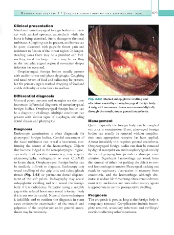
Guttural pouch mycosis and strangles are the most Fig. 3.63 Marked subepiglottic swelling and
important differential diagnoses of nasopharygneal ulceration caused by an oropharyngeal foreign body.
foreign bodies. Oropharyngeal foreign bodies can A twig with numerous thorns was removed digitally
be a diagnostic challenge. Multiple conditions can through the mouth, under general anaesthesia.
present with similar signs of dysphagia, including
dental disease and pharyngitis. Management
Quite frequently the foreign body can be coughed
Diagnosis out prior to examination. If not, pharyngeal foreign
Endoscopic examination is often diagnostic for bodies can usually be removed without complica-
pharyngeal foreign bodies. Careful assessment of tion once appropriate restraint has been applied.
the nasal turbinates can reveal a laceration, con- Almost invariably this requires general anaesthesia.
firming the source of the haemorrhage. Objects Oropharyngeal foreign bodies can then be removed
that become lodged in the retropharyngeal region, by digital manipulation and nasopharyngeal ones by
especially if of wooden consistency, may require the use of grasping forceps under endoscopic visu-
ultrasonography, radiography or even CT/MRI alisation. Significant haemorrhage can result from
to locate them. Oropharyngeal foreign bodies can the removal of either but packing the defect to con-
be similarly difficult to diagnose. Endoscopy may trol haemorrhage is unwise. Pharyngeal packing can
reveal swelling of the epiglottis and subepiglottic result in respiratory obstruction in recovery from
tissues (Fig. 3.63) or permanent dorsal displace- anaesthesia, and the haemorrhage, although dra-
ment of the soft palate. Radiography may reveal matic, is seldom life threatening. Once removed treat-
subepiglottic swelling and will reveal the foreign ment with antibiotics and anti-inflammatory agents
body if it is radiodense. Palpation using a suitable is appropriate to control postoperative swelling.
gag in the sedated horse may reveal a foreign body
if it is not too far caudal. None of these techniques Prognosis
is infallible and to confirm the diagnosis in some The prognosis is good as long as the foreign body is
cases endoscopic examination of the mouth and completely removed. Complications include incom-
palpation of the oropharynx under general anaes- plete removal, secondary infections and swellings/
thesia may be necessary. reactions affecting other structures.