
Page 448 - Fluid, Electrolyte, and Acid-Base Disorders in Small Animal Practice
P. 448
CHAPTER • 18
Fluid and Electrolyte
Disturbances In Gastrointestinal
and Pancreatic Disease
Joao Felipe de Brito Galvao, Kenneth W. Simpson, and Nichole Birnbaum
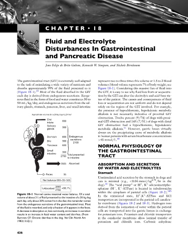
The gastrointestinal tract (GIT) is extremely well adapted represent two to three times this volume or 1.5 to 2 blood
to the task of assimilating a wide variety of nutrients and volumes (blood volume represents 7% of body weight; see
absorbs approximately 99% of the fluid presented to it Figure 18-1). Considering this massive flux of fluid into
(Figure 18-1). 15 Most of the fluid absorbed in the GIT the GIT, it is easy to see why fluid loss from or sequestra-
each day is derived from endogenous secretions. Exoge- tion by the GITcan alter the electrolyte and acid-base sta-
nous fluid in the form of food and water constitutes 30 to tus of the patient. The causes and consequences of fluid
50 mL/kg/day, and endogenous secretions from the sal- loss or sequestration are not uniform and do not depend
ivary glands, stomach, pancreas, liver, and small intestine solely on the region of the GIT involved. For example,
the presence of hypochloremic, hypokalemic metabolic
Approximate volumes for a 20-kg dog mL/24 hrs* alkalosis is not necessarily indicative of proximal GIT
obstruction. Twelve percent (9/74) of dogs with proxi-
Oral 600
intake mal GIT obstruction and 14% (7/51) of dogs with distal
GIT obstruction had a hypochloremic, hypokalemic
Salivary
300 11
glands metabolic alkalosis. However, gastric losses virtually
always are the precipitating cause of metabolic alkalosis
Stomach 600 Endogenous in human patients with serum bicarbonate concentrations
secretions >45 mmol/L. 40
Bile 300
2100
Pancreas 600 NORMAL PHYSIOLOGY OF
Small 300 THE GASTROINTESTINAL
intestine Total presented
2700 TRACT
to intestine
2665 Absorbed ABSORPTION AND SECRETION
OF WATER AND ELECTROLYTES
Stomach
Feces 35
Unstimulated acid secretion by the stomach in dogs and
Net balance 600–35=565 0.75
cats is minimal (e.g., <0.04 mmol/kg /hr in the
þ
þ
dog). 45 The “acid pump” or H ,K -adenosinetripho-
2665
þ
þ
%Absorbed =98.7% sphatase (H ,K -ATPase) is located in tubulovesicles
2700 105
Figure 18-1 Normal canine intestinal water balance. Of a total within the cytoplasm of parietal cells (Figure 18-2).
þ
þ
volume of about 3 L of fluid presented to the intestine of a 20-kg dog In the stimulated state, H ,K -ATPase and KCl
each day, only about 20% comes from the diet; the remainder comes transporters are incorporated in the parietal cell canalicu-
from the endogenous secretions of the gastrointestinal tract. Most lar membrane (Figures 18-2 and 18-3). Hydrogen ions
of this fluid is resorbed, and only a fraction of it appears in the feces. derived from the ionization of water within the parietal
A decrease in absorption or, less commonly, an increase in secretion cells are transported into the gastric lumen in exchange
results in an increase in fecal water content and diarrhea. (From for potassium ions. Potassium and chloride transporters
Burrows CF. Chronic diarrhea in the dog. Vet Clin North Am in the canalicular membrane allow luminal transfer of
1983;13:521.) potassium and chloride ions. Carbonic anhydrase
436