
Page 473 - Fluid, Electrolyte, and Acid-Base Disorders in Small Animal Practice
P. 473
Fluid, Electrolyte, and Acid-Base Disturbances in Liver Disease 461
and permeability characteristics and augments platelet causing extracorporeal albumin loss and an acute-phase
aggregation, which may predispose to thromboembolic response (e.g., decreased albumin synthesis, increased
complications. 231 The clinical implication of a lower transcapillary loss).
reduced/oxidized albumin ratio lies in its relationship Absolute hyperalbuminemia is exceedingly rare, but
to oxidative stress imposed by low thiol substrate has been reported in one dog and one human patient with
availability. hepatocellular carcinoma. Hyperalbuminemia was
Numerous factors influence serum albumin concentra- hypothesized to be a consequence of increased synthesis
tion (see Figure 19-3). Modest hypoalbuminemia may of albumin by malignant hepatocytes or due to decreased
reflect reduced albumin synthesis or enhanced catabo- negative feedback from impaired hepatocellular
lism, but these usually are slow in onset. Protein catabo- osmoreceptivity. 56,157
lism caused by illness usually spares albumin and targets
muscle. The acute-phase response to tissue injury Globulins
enhances transcapillary escape of albumin and may reduce The plasma globulin concentration represents many
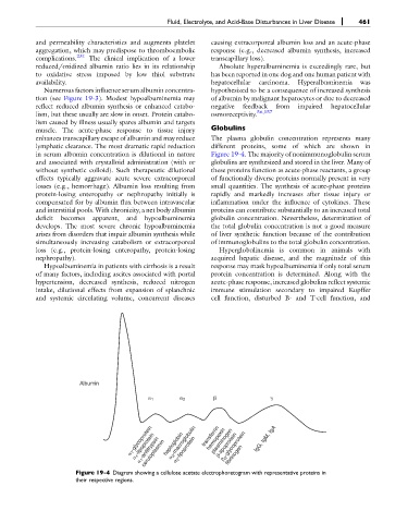
lymphatic clearance. The most dramatic rapid reduction different proteins, some of which are shown in
in serum albumin concentration is dilutional in nature Figure 19-4. The majority of nonimmunoglobulin serum
and associated with crystalloid administration (with or globulins are synthesized and stored in the liver. Many of
without synthetic colloid). Such therapeutic dilutional these proteins function as acute-phase reactants, a group
effects typically aggravate acute severe extracorporeal of functionally diverse proteins normally present in very
losses (e.g., hemorrhage). Albumin loss resulting from small quantities. The synthesis of acute-phase proteins
protein-losing enteropathy or nephropathy initially is rapidly and markedly increases after tissue injury or
compensated for by albumin flux between intravascular inflammation under the influence of cytokines. These
and interstitial pools. With chronicity, a net body albumin proteins can contribute substantially to an increased total
deficit becomes apparent, and hypoalbuminemia globulin concentration. Nevertheless, determination of
develops. The most severe chronic hypoalbuminemia the total globulin concentration is not a good measure
arises from disorders that impair albumin synthesis while of liver synthetic function because of the contribution
simultaneously increasing catabolism or extracorporeal of immunoglobulins to the total globulin concentration.
loss (e.g., protein-losing enteropathy, protein-losing Hyperglobulinemia is common in animals with
nephropathy). acquired hepatic disease, and the magnitude of this
Hypoalbuminemia in patients with cirrhosis is a result response may mask hypoalbuminemia if only total serum
of many factors, including ascites associated with portal protein concentration is determined. Along with the
hypertension, decreased synthesis, reduced nitrogen acute-phase response, increased globulins reflect systemic
intake, dilutional effects from expansion of splanchnic immune stimulation secondary to impaired Kupffer
and systemic circulating volume, concurrent diseases cell function, disturbed B- and T-cell function, and
Albumin
1 2
2 -macroglobulin
1 -glycoprotein haptoglobin transferrin plasminogen fibrinogen IgG, IgM, IgA
hemopexin
2 -glycoprotein
-lipoprotein
1 -lipoprotein
2 -lipoprotein
1 -antitrypsin
ceruloplasmin
Figure 19-4 Diagram showing a cellulose acetate electrophoretogram with representative proteins in
their respective regions.