
Page 490 - Fluid, Electrolyte, and Acid-Base Disorders in Small Animal Practice
P. 490
478 FLUID THERAPY
90
(n = 30)
80
Cirrhotic dogs (%) 60
70
50
40
30
20
10
0
↓pH ↑pH ↑CO ↑SID ↓SID ↑Na ↓Na ↑Cl ↓Cl ↑XA ↑XA ↑Alb ↓Alb ↑Phos ↓Phos ↑AG ↑AG
2 ↓CO 2
Adjusted Adjusted
Acid-base disturbance parameters
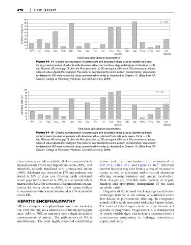
Figure 19-14 Graphic representation of parameters and calculated values used to identify acid-base
derangements (number of patients with abnormal values) derived from dogs with hepatic cirrhosis (n ¼ 30).
Alb, Albumin; AG, anion gap; Cl, chloride; Phos, phosphorus; SID, strong ion difference; XA, unmeasured anions;
Adjusted, value adjusted for change in free water as represented by serum sodium concentration. Values used
to determine SID were calculated using conventional formulas as described in Chapter 13. (Data from SA
Center: College of Veterinary Medicine, Cornell University, 2004).
100
(n = 23)
90
Lipidosis cats (%) 70
80
60
50
40
30
20
10
0
↓pH ↑pH ↑CO 2 ↓CO 2 ↑SID ↓SID ↑Na ↓Na ↑Cl ↓Cl ↑XA ↑XA ↑Alb ↓Alb ↑Phos ↓Phos ↑AG ↑AG
Adjusted Adjusted
Acid-base disturbance parameters
Figure 19-15 Graphic representation of parameters and calculated values used to identify acid-base
derangements (number of patients with abnormal values) derived from cats with severe HL (n ¼ 23).
Alb, Albumin; AG, anion gap; Cl, chloride; Phos, phosphorus; SID, strong ion difference; XA, unmeasured anions;
Adjusted, value adjusted for change in free water as represented by serum sodium concentration. Values used
to determine SID were calculated using conventional formulas as described in Chapter 13. (Data from SA
Center: College of Veterinary Medicine, Cornell University, 2004).
these cats may include metabolic alkalosis associated with factors and their mechanisms are summarized in
hypochloremia (74%) and hypoalbuminemia (48%), and Box 19-2, Table 19-3, and Figure 19-16. 47 Abnormal
metabolic acidosis associated with unmeasured anions cerebral function may arise from a variety of neuroactive
(96%). Alkalemia was detected in 17% and acidemia was toxins, as well as functional and structural alterations
found in 26% of these cats. Conventionally calculated affecting neurotransmission and energy metabolism.
anion gaps were abnormal in 39%, but abnormal values Most changes are reversible with recovery of hepatic
increasedto52%aftercorrectionofserumsodiumconcen- function and appropriate management of the acute
tration for water excess or deficit. Low serum sodium metabolic crisis.
concentration (water excess) was found in 57% of cats with Diagnosis of HE is based on clinical signs and clinico-
severe HL. pathologic features in the setting of confirmed severe
liver disease or portosystemic shunting. In companion
HEPATIC ENCEPHALOPATHY animals, HE is rarely associated with acute hepatic failure.
HE is a complex neurophysiologic syndrome involving The onset of clinical signs can be acute or chronic and
the CNS that implies a critical loss of functional hepatic episodic or progressive. Progressive HE is characterized
mass (65% to 70%) or extensive hepatofugal circulation by widely variable signs that include a decreased level of
(portosystemic shunting). The pathogenesis of HE is consciousness progressing to lethargy, somnolence,
multifactorial. The most highly suspected contributing stupor, and coma.