
Page 1084 - Adams and Stashak's Lameness in Horses, 7th Edition
P. 1084
1050 Chapter 10
VetBooks.ir
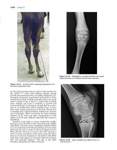
Figure 10.19. Radiograph of a newborn foal that has a carpal
valgus deformity due to delayed cuboidal bone ossification.
Figure 10.18. Young foal with a windswept appearance to the
tarsi due to ligamentous laxity.
for the foal is not the same as correct conformation for
the adult. 2,3,16,26 Foals will undergo changes during
growth and maturation that can rapidly change the con
formation. In general, examination of the foal for ALDs
should start as close to birth as possible, then every week
until 1 month of age as this is a critical time in which
many changes can occur in response to growth and
exercise demands. Examinations should then be contin
ued on a monthly basis until 6 months of age. A com
plete exam entails examination of the limb of interest in
a standing and a flexed position, followed by exercise
and radiographs. When examining the limb standing,
rotation of the limb can make interpretation of the
degree of ALDs more difficult, especially those distal to
the fetlock.
Rotation of the limb or torsion around the weight‐
bearing axis of the limb is often associated and coupled
with ALDs. Therefore, when examining a limb for ALDs,
it is very important to be directly lined up in front of the
limb and not just in front of the foal (Figure 10.22).
Being directly lined up in front of the limb will give a
more accurate assessment of the ALD, especially when
there is a rotational component to the limb as well. Most
foals have some degree of external (toed‐out) rotation
that will generally improve with age as the chest Figure 10.20. Septic cuboidal bones (black arrows) in a
broadens and pushes the elbow outward. 1‐month‐old colt.