
Page 1086 - Adams and Stashak's Lameness in Horses, 7th Edition
P. 1086
VetBooks.ir
A B
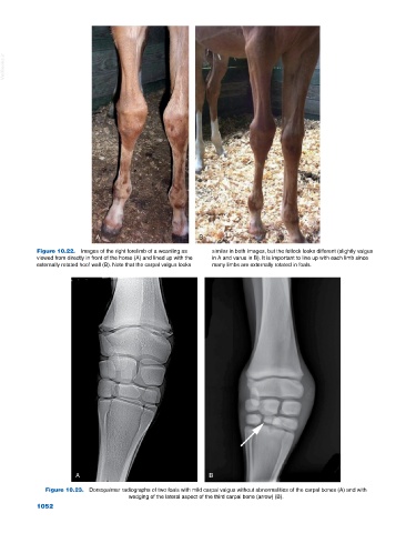
Figure 10.22. Images of the right forelimb of a weanling as similar in both images, but the fetlock looks different (slightly valgus
viewed from directly in front of the horse (A) and lined up with the in A and varus in B). It is important to line up with each limb since
externally rotated hoof wall (B). Note that the carpal valgus looks many limbs are externally rotated in foals.
A B
Figure 10.23. Dorsopalmar radiographs of two foals with mild carpal valgus without abnormalities of the carpal bones (A) and with
wedging of the lateral aspect of the third carpal bone (arrow) (B).
1052