
Page 1091 - Adams and Stashak's Lameness in Horses, 7th Edition
P. 1091
VetBooks.ir
A B
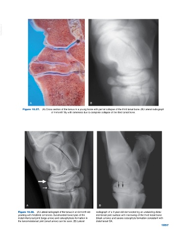
Figure 10.27. (A) Cross section of the tarsus in a young horse with partial collapse of the third tarsal bone. (B) Lateral radiograph
of 4‐month filly with lameness due to complete collapse of the third tarsal bone.
A B
Figure 10.28. (A) Lateral radiograph of the tarsus in an 8‐month‐old radiograph of a 2‐year‐old demonstrating an undulating distal
yearling with hindlimb lameness. Subchondral bone lysis of the intertarsal joint surface with narrowing of the third tarsal bone
distal intertarsal joint (large arrow) and osteophytosis formation in (black arrows) and severe osteophyte formation consistent with
the tarsometatarsal joint (small arrow) can be seen. (B) Lateral distal tarsal OA.
1057