
Page 1095 - Adams and Stashak's Lameness in Horses, 7th Edition
P. 1095
Lameness in the Young Horse 1061
be used with caution because they may produce excessive
tension on the laminae. 10,27
VetBooks.ir can often be necessary to help offset the pain that is
While NSAID treatment in young foals is not ideal, it
produced from the additional therapies such as splinting.
It is important to have the foals be comfortable enough
to stand as they will improve quicker than if then remain
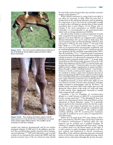
recumbent due to pain (Figure 10.33). Please refer to
Chapter 8 for more information on NSAID therapy.
Generally, it is recommended to use gastroprotectants
while foals are being administered NSAIDs.
Oxytetracycline remains a common treatment for foals
with congenital flexural deformities. 1,2,10,19,23 Clinically, it
appears to be more effective on congenital than acquired
flexural deformities, but can be used for both types. The
use of oxytetracycline was first described in 1985 by Lokai
and again in 1992 by the same author. 19,20 All foals in the
latter study (n = 123) were treated either once or twice
with 3 g of oxytetracycline intravenously (undiluted), and
Figure 10.31. Foal with a tarsal and fetlock flexural deformity of no external splinting was used. A 94% overall success rate
the left hindlimb as well as bilateral carpal and forelimb fetlock was reported with the forelimbs responding better (98%)
19
flexural deformities. than the hindlimbs (75%). Oxytetracycline’s mechanism
of action has been hypothesized to result from chelation of
calcium in muscle or to decrease the expression of the con
tractile protein α‐smooth muscle actin. 14,19 A recent study
found that myofibroblasts made up most of the cells in the
distal check ligament and the DDFT in normal foals, sup
porting this rationale. Oxytetracycline was also found to
14
inhibit normal collagen organization in equine myofibro
blasts through a matrix metalloproteinase‐1 mechanism.
5
However it works, oxytetracycline appears to be an effec
tive method to obtain a short‐term moderate decrease in
metacarpophalangeal (MCP) joint angle in newborn foals.
Two controlled studies have documented changes in MCP
joint angles following treatment with oxytetracycline. 16,21
A significant decrease in mean MCP joint angles at rest
was observed in foals 24 hours after treatment, and these
angles returned to pretreatment value by 96 hours. In
21
another study, maximum MCP joint angles that occurred
during the stance phase of the stride at a walk and range
of joint motion were significantly increased in treated
foals compared with controls. 16
Currently, 1–3 g (or 44 mg/kg) of oxytetracycline
diluted in a small amount of saline IV is recommended
for the average‐sized foal. 12,18 The 44 mg/kg IV dose was
used in one experimental study with good effects and no
alterations in blood chemistry values. It is generally
21
ideal to perform this treatment within the first few weeks
of life. One or two repeat treatments may be given, but
the risk of renal problems should be considered with
multiple dosing or overdosing. 10,37 The authors usually
combine splinting with IV oxytetracycline treatment,
especially in moderate to severe cases of congenital flex
ural deformities.
Figure 10.32. Fluid swelling at the lateral aspect of the left Splints may be made from PVC pipe tubing or fiber
carpus (arrow) in a 3‐day‐old foal that is characteristic of a rupture glass cast material. The PVC pipe can be bent with heat
of the common digital extensor tendon. This condition can be to whatever conformation is desired while the cast
unilateral as in this foal or bilateral.
material can be molded over a padded bandage. In
addition, IV simplex tubing has also been used, acting
18
carpus) may improve spontaneously and do not require as a rubber band over the dorsal aspect of the limb. It
treatment (Figures 10.29B and 5.6). In addition, most fet is often easiest to apply bandages and splints with the
lock flexural deformities usually respond well to bandag foal sedated and positioned in lateral recumbency.
ing and splinting. 2,10,23 DIP joint deformities may respond Regardless of the method, splinting increases tensional
to glue‐on toe extensions that stretch the DDFT and also forces to the flexor units during weight‐bearing, which
prevent excessive wear of the toe. However, these should induces flexor relaxation through the inverse myotatic