Page 1099 - Adams and Stashak's Lameness in Horses, 7th Edition
P. 1099

Lameness in the Young Horse  1065


                                                                 lameness should cause wariness of other problems in the
                                                                 limb. Dishing or concavity of the dorsal hoof wall may
  VetBooks.ir                                                    the dorsal laminae may lead to seedy toe and toe
                                                                 occur in chronic cases (Figure 10.35B), and tearing of
                                                                 abscesses.
                                                                   The severity of the deformity may be subdivided into
                                                                 stages I and II based on the visual position of the dorsal
                                                                 hoof wall. 23,24  Stage I contracture is when the dorsal
                                                                 hoof wall does not go beyond vertical (Figure 10.35A),
                                                                 and stage II is when the dorsal surface of the hoof passes
                                                                 beyond vertical (Figure  10.38).  The more severe the
                                                                 deformity, the more shortening of the musculotendinous
                                                                 unit, and the more aggressive treatment should be. With
                                                                 severe contracture, pathologic changes may develop in
                                                                 the joint capsule and other tissues of the coffin joint,
                                                                 making permanent correction less likely. 13,23  With chro­
                                                                 nicity, more changes can happen to the hoof capsule due
                                                                 to the pressure being placed on the toe. The degree of
                                                                 concavity at the toe and discrepancy in growth rings
                                                                 from dorsal to palmar have been categorized into differ­
                                                                 ent stages. 8,27
                                                                   Flexural deformity of the MCP joint has been classi­
                                                                 cally referred to as contracture of the superficial digital
                                                                 flexor tendon (SDFT). It is characterized by dorsal
                                                                 knuckling of the fetlock with the hoof itself remaining
                                                                 in normal alignment (Figures  10.36 and 10.37).  The
                                                                 term SDFT contracture is a gross oversimplification
                                                                 because the SDFT, DDFT, both the SDFT and DDFT,
                                                                 and the suspensory ligament in chronic cases may the
                                                                                         2
                                                                 involved with the deformity.  Early in the condition, the
                                                                 fetlock and pastern may begin to appear more upright
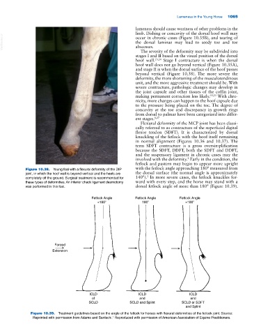
             Figure 10.38.  Young foal with a flexural deformity of the DIP   with the fetlock angle approaching 180° measured from
             joint, in which the hoof wall is beyond vertical and the heels are   the dorsal surface (the normal angle is approximately
                                                                      2
             completely off the ground. Surgical treatment is recommended for   140°).  In more severe cases, the fetlock knuckles for­
             these types of deformities. An inferior check ligament desmotomy   ward with every step, and the horse may stand with a
             was performed in this foal.                         dorsal fetlock angle of more than 180° (Figure 10.39).

                                            Fetlock Angle        Fetlock Angle       Fetlock Angle
                                               <180°                180°                >180°


                           Forced
                          Extension


                                           ICLD                   ICLD                    ICLD
                                            of                    and                     and
                                           SCLD               SCLD and Splint         SCLD or SDFT
                                                                                        and Splint
               Figure 10.39.  Treatment guidelines based on the angle of the fetlock for horses with flexural deformities of the fetlock joint. Source:
                                                      1
                Reprinted with permission from Adams and Santschi.  Reproduced with permission of American Association of Equine Practitioners.
   1094   1095   1096   1097   1098   1099   1100   1101   1102   1103   1104