
Page 1100 - Adams and Stashak's Lameness in Horses, 7th Edition
P. 1100
1066 Chapter 10
As with flexural deformities of the DIP joint, when the
deformity goes beyond vertical (constant dorsal knuck
VetBooks.ir required. In chronic cases, the suspensory ligament
ling of the fetlock), more aggressive treatment is usually
becomes involved, fibrosis of the joint capsule may
occur, and osteoarthritic changes may develop in the
fetlock joint. 23
Diagnosis
A tentative diagnosis of flexural deformities of the
DIP or MCP joints usually can be made based on the
characteristic foot and limb conformation. Flexural
deformities of the DIP joint only involve the DDFT.
Determining which soft tissue structure(s) is involved
with deformities of the MCP joint is more difficult.
Since these acquired deformities can developed second
ary to a painful focus, such as physitis, it is important to
identify any potential sources of pain, as without elimi
nating those, it will be very difficult to treat the flexural
deformity.
The horse should be examined from a distance in its
natural environment and then on flat ground to deter
mine how much the limb can straighten while moving
or standing flat. Standing palpation should then be
performed to determine the presence of any asym
metries that could be related to physitis or joint effu
sion (possible OCD). Similar to congenital cases, the
clinician can attempt to manually straighten the limb
while simultaneously palpating the flexor surface of
the limb to see if any specific structure becomes more
taught. For instance, the DDFT, SDFT, and the suspen
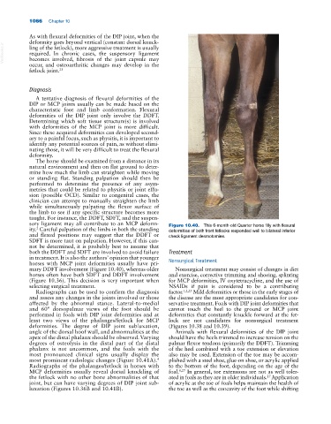
sory ligament may all contribute to an MCP deform Figure 10.40. This 6‐month‐old Quarter horse filly with flexural
ity. Careful palpation of the limbs in both the standing deformities of both front fetlocks responded well to bilateral inferior
2
and flexed positions may suggest that the DDFT or check ligament desmotomies.
SDFT is more taut on palpation. However, if this can
not be determined, it is probably best to assume that
both the DDFT and SDFT are involved to avoid failure Treatment
in treatment. It is also the authors’ opinion that younger Nonsurgical Treatment
horses with MCP joint deformities usually have pri
mary DDFT involvement (Figure 10.40), whereas older Nonsurgical treatment may consist of changes in diet
horses often have both SDFT and DDFT involvement and exercise, corrective trimming and shoeing, splinting
(Figure 10.36). This decision is very important when for MCP deformities, IV oxytetracycline, and the use of
selecting surgical treatment. NSAIDs if pain is considered to be a contributing
Radiographs can be used to confirm the diagnosis factor. 1,2,23 Mild deformities or those in the early stages of
and assess any changes in the joints involved or those the disease are the most appropriate candidates for con
affected by the abnormal stance. Lateral‐to‐medial servative treatment. Foals with DIP joint deformities that
and 60° dorsopalmar views of the foot should be cannot touch the heel to the ground or MCP joint
performed in foals with DIP joint deformities and at deformities that constantly knuckle forward at the fet
least two views of the phalanges/fetlock for MCP lock are not candidates for nonsurgical treatment
deformities. The degree of DIP joint subluxation, (Figures 10.38 and 10.39).
angle of the dorsal hoof wall, and abnormalities at the Animals with flexural deformities of the DIP joint
apex of the distal phalanx should be observed. Varying should have the heels trimmed to increase tension on the
degrees of osteolysis in the distal part of the distal palmar flexor tendons (primarily the DDFT). Trimming
phalanx is not uncommon, and the foals with the of the heel combined with a toe extension or elevation
most pronounced clinical signs usually display the also may be used. Extension of the toe may be accom
most prominent radiologic changes (Figure 10.41A). plished with a steel shoe, glue‐on shoe, or acrylic applied
4
Radiographs of the phalanges/fetlock in horses with to the bottom of the foot, depending on the age of the
MCP deformities usually reveal dorsal knuckling of foal. 8,27 In general, toe extensions are not as well toler
the fetlock with no other bone abnormalities of that ated in foals as they are in older individuals. Application
27
joint, but can have varying degrees of DIP joint sub of acrylic at the toe of foals helps maintain the health of
luxation (Figures 10.36B and 10.41B). the toe as well as the concavity of the foot while shifting