
Page 1098 - Adams and Stashak's Lameness in Horses, 7th Edition
P. 1098
1064 Chapter 10
However, the relationship between age and the type of flex deformities of both the DIP and MCP joints develop subse
ural deformity is not absolute. quent to trauma to the elbow region.
VetBooks.ir Etiology produce flexural deformities is still uncertain. Suggested
The mechanism by which the suggested risk factors
hypotheses include the failure of tendons and ligaments
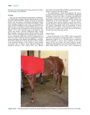
The cause of acquired flexural deformities is multifacto to develop at the same rate as bone lengthening and a
rial. Historically, acquired flexural deformities have been discrepancy between bone growth and the capacity for
grouped within the DOD complex because the potential lengthening of the check ligaments. 13,23 Flexor muscles
causes and risk factors (overfeeding, nutritional imbal are stronger than extensor muscles, and the foal conse
ances, trauma, and genetics) are similar. In addition, other quently develops a flexural deformity. These theories are
23
types of DOD conditions such as physitis and OCD can not entirely compatible with our knowledge of bone
often be seen concurrently in these animals and may actu growth since the majority of bone growth occurs early in
ally contribute to the development of flexural deformities life (first few months) and many flexural deformities
(Figure 10.37). This is because pain in the limb for any develop later than that time period.
reason may initiate a flexion withdrawal reflex causing
flexor muscle contraction and an altered position of the Clinical Signs
joint. Chronic unweighting of the leg also may contribute
23
to foot contracture, high heels, and the development of a Flexural deformity of the DDFT centers around the
club foot. Lack of exercise has been suggested to prevent DIP joint and results in a raised heel with a “club foot”
proper stretching of the tendons and ligaments, contribut appearance (Figure 10.35). The heel may be completely
ing to limb contracture. Severe trauma to a flexor tendon off the ground in severe cases (walking on the toe;
23
or its associated muscles is also known to cause tendon Figure 10.38), but usually the heels maintain contact
contracture due to fibrous tissue deposited during the with the ground and grow excessively long (Figure 10.35).
13
reparative process. The authors have seen flexural Most foals should not be lame, and a pronounced
Figure 10.37. This yearling Quarter horse filly had flexural deformities of both front fetlocks and subchondral cystic lesions in both stifles.