
Page 1088 - Adams and Stashak's Lameness in Horses, 7th Edition
P. 1088
1054 Chapter 10
the advantages of HCTP + PS over TPB techniques are the implants must be removed or the limb will over cor
that the procedure is easy to perform, has minimal rect. Limb growth usually returns to normal following
VetBooks.ir and the limb does not overcorrect. In addition, the tion has been seen following removal of the transphy
implant removal. However, continued growth retarda
complications, and is less expensive than TPB procedures
seal screw, and this should be considered when deciding
majority of horses can perform well after surgery,
although racing performance has been shown to be when and where to use this technique. Other considera
reduced if HCTP+PS is performed at two or more tions for a transphyseal screw are the potential for the
23
anatomic locations. Despite these benefits, HCTP+PS development of a physitis that results in metaphyseal
appears to be most useful for mild to moderate cases of collapse (creates an angular deformity in the opposite
9
ALD that have not responded to conservative treatment. direction) when used for carpal ALDs. Due to this and
Foals with severe ALD at any location are best treated the fact that use of a transphyseal screw increased the
with TPB. risk of physitis that required further treatment, sur
Retardation of endochondral ossification on the con geons should carefully weigh the pros and cons of using
9
vex side of the deformity can be accomplished with vari a single screw in the carpus. The advantages of the TPB
ous TPB procedures. 10,13,18,20,22,33 The bridges may consist procedures are that severe deformities can be corrected
of staples, 10,18 screws and wires, 4,22 a small plate spanning quickly and most surgeons feel that the results are more
the growth plate, or a transphyseal lag or positional reliable than HCTP + PS. The disadvantages include
18
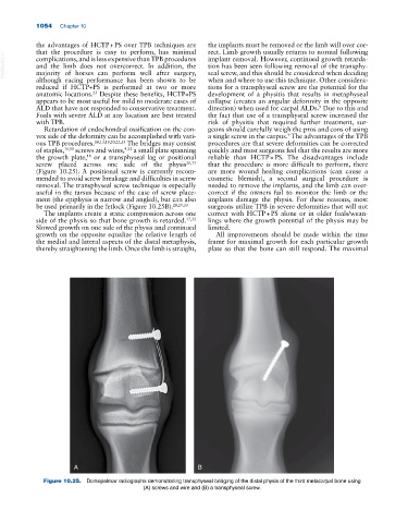
screw placed across one side of the physis 20,33 that the procedure is more difficult to perform, there
(Figure 10.25). A positional screw is currently recom are more wound healing complications (can cause a
mended to avoid screw breakage and difficulties in screw cosmetic blemish), a second surgical procedure is
removal. The transphyseal screw technique is especially needed to remove the implants, and the limb can over
useful in the tarsus because of the ease of screw place correct if the owners fail to monitor the limb or the
ment (the epiphysis is narrow and angled), but can also implants damage the physis. For these reasons, most
be used primarily in the fetlock (Figure 10.25B). 20,29,33 surgeons utilize TPB in severe deformities that will not
The implants create a static compression across one correct with HCTP + PS alone or in older foals/wean
side of the physis so that bone growth is retarded. 17,32 lings where the growth potential of the physis may be
Slowed growth on one side of the physis and continued limited.
growth on the opposite equalize the relative length of All improvements should be made within the time
the medial and lateral aspects of the distal metaphysis, frame for maximal growth for each particular growth
thereby straightening the limb. Once the limb is straight, plate so that the bone can still respond. The maximal
A B
Figure 10.25. Dorsopalmar radiographs demonstrating transphyseal bridging of the distal physis of the third metacarpal bone using
(A) screws and wire and (B) a transphyseal screw.