
Page 1165 - Adams and Stashak's Lameness in Horses, 7th Edition
P. 1165
Foot Care and Farriery 1131
VetBooks.ir
A B
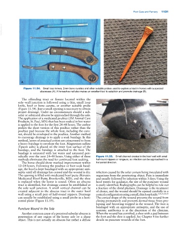
Figure 11.54. Small loop knives, 2 mm bone curettes and other suitable probes used to explore a tract in horses with suspected
abscesses (A). A horseshoe nail also makes an excellent tool to establish and promote drainage (B).
The offending tract or fissure located within the
sole–wall junction is followed using a thin, small loop
knife, hoof or bone curette, or another suitable probe
(Figure 11.54). Just a small opening is necessary to obtain
proper drainage. Under no circumstances should a sub
solar or submural abscess be approached through the sole.
The application of a medicated poultice (3M Animal Care
Products, St. Paul, MN) that has been soaked in hot water
is applied to the foot for the first 24–48 hours. The author
prefers the sheet version of this poultice rather than the
poultice pad because the whole foot, including the coro
net, should be enveloped in the poultice. Another method
to encourage drainage is to apply a soak bandage. In this
method, layers of practical cotton are crisscrossed to form
a heavy bandage to envelope the foot. Magnesium sulfate
(Epsom salts) is placed on the inner foot surface of the
bandage, and the bandage is attached to the foot. The
bandage is saturated with hot water and saturated peri
odically over the next 24–48 hours. Using either of these Figure 11.55. Small channel created in the hoof wall with small
methods eliminates the need for continued foot soaking. half‐round nippers or rongeurs, so infection can be approached in a
The horse should show marked improvement within horizontal direction.
12–24 hours. Following the poultice or foot soak band
age, the hoof is kept bandaged with an appropriate anti
septic until all drainage has ceased and the wound is dry. infection caused by the solar corium being inoculated with
The opening is filled with medicated hoof putty (Keratex organisms from the penetrating object. Pain is immediate
Medicated Hoof Putty, Brookeville, MD), and the shoe and usually followed by infection within 3 days. Using the
is replaced when the horse is sound. Often an abscess hoof testers for guidance, the site of the puncture wound
tract is identified, but drainage cannot be established at is easily identified. Radiographs can be helpful to rule out
the sole–wall junction. A small vertical channel can be a fracture of the distal phalanx. Drainage is the treatment
created adjacent to the abscess tract in the outer hoof of choice, and the wound should be opened carefully in a
wall using a small pair of half‐round nippers. Drainage conical configuration using a small, thin loop knife. 9,40,42,43,45
can usually be established using a small probe in a hori The conical shape of the wound prevents the wound from
zontal plane (Figure 11.55). closing prematurely and prevents dermal tissue from pro
lapsing and becoming trapped in the wound. The foot is
Puncture Wound in the Sole bandaged with an appropriate antiseptic, and the use of
systemic antibiotics is at the discretion of the clinician.
Another common cause of a perceived subsolar abscess is When the wound has cornified, a shoe with a pad between
penetration of any region of the horny sole by a sharp the foot and the shoe is applied. See Chapter 4 for further
object. This is not actually an abscess but rather a diffuse details on puncture wounds of the foot.