
Page 1164 - Adams and Stashak's Lameness in Horses, 7th Edition
P. 1164
1130 Chapter 11
Treatment for sole pressure is to concave the inner foot pressures on the affected heel by unloading the heel with
surface of the shoe to relieve the pressure on the sole or, some form of bar shoe.
VetBooks.ir the inner foot surface of the shoe concaved or deep seated Hoof Abscesses
in the case of the horse without shoes, apply a shoe with
to raise the solar surface of the foot off the ground.
The last form of bruising is the so‐called corn, which Hoof abscess, a localized accumulation of purulent
is focal hemorrhage that occurs in the angle of the sole exudate located between the germinal and keratinized
at the heels between the hoof wall and the bar (seat of layers of the epithelium, most commonly subsolar or
the corn). The hemorrhage results from trauma, origi submural, is probably the most common cause of acute
nates from the dermal lamellae, and appears as a small, lameness. Entry is either through a break or fissure in
42
focal, red discoloration in the seat of the corn. It may be the sole–wall junction (white line), a misplaced nail, or a
present in one or both heels. There may be a marked puncture wound somewhere in the solar surface of the
hoof tester response, and the source of pain can be con foot. Organisms also may enter the foot by way of a
firmed using unilateral local anesthesia. Confusing the full‐thickness hoof wall defect or crack.
diagnosis are abscess, hoof wall separation, and fracture Most horses with a foot abscess show an acute onset
of the bars. Treatment of corns involves removing the of lameness. The degree of lameness varies from being
subtle in the early stages to non‐weight‐bearing. The
digital pulse is usually bounding, and with careful obser
vation, unless the abscess is in the middle of the toe, the
intensity of the digital pulse is much stronger on the side
of the foot where the abscess is located. If the abscess is
long standing, there may be soft tissue swelling in the
pastern or above the fetlock on the side of the limb cor
responding to the side of the foot where the abscess is
located. The site of pain can be localized to a small focal
area through the careful use of hoof testers.
The most important aspect of treating a subsolar/
submural abscess is establishing drainage. The opening
should be sufficient to allow drainage but not so exten
sive as to create further damage to the hoof capsule.
When pain is localized with hoof testers, a small tract
or fissure will commonly be found in the sole–wall
junction (white line) (Figure 11.53A and B). The wound
or point of entry may not always be visible, because
some areas of the foot such as the white line are some
Figure 11.52. Foot with a rim pad placed between the solar what elastic and wounds in this area tend to close. In
surface of the foot and the shoe, which raises the sole higher off the this case, a suitable poultice should be applied to the
ground. The weight of the horse forces the sole through the foot daily in an attempt to soften the affected area, and
branches of the shoe toward the ground. eventually a tract will become obvious.
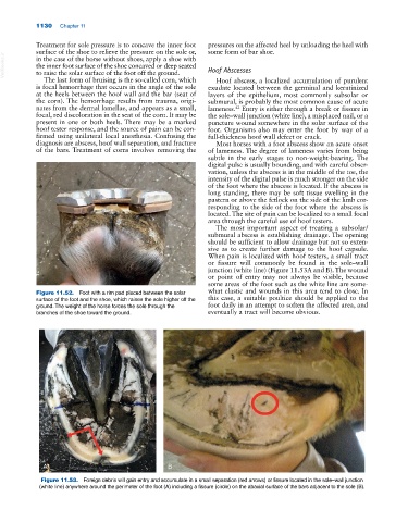
A B
Figure 11.53. Foreign debris will gain entry and accumulate in a small separation (red arrows) or fissure located in the sole–wall junction
(white line) anywhere around the perimeter of the foot (A) including a fissure (circle) on the abaxial surface of the bars adjacent to the sole (B).