
Page 1159 - Adams and Stashak's Lameness in Horses, 7th Edition
P. 1159
Foot Care and Farriery 1125
VetBooks.ir
A B
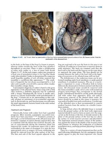
Figure 11.47. (A) Thrush. Note the deterioration of the frog that is recessed below ground surface of foot. (B) Severe canker. Note the
proliferation of the diseased horn.
to the body or the base of the frog if a fissure is present, frog are explored to be sure the horn in this area is not
whereas canker invades the horn of the frog anywhere involved; all underrun or loose horn is removed down to
throughout its structure. There is often a proliferation solid structures. The heels are trimmed such that the
of tissue with canker vs. a loss of tissue with thrush hoof wall at the heels and the frog are on the same plane
(Figure 11.47). In the early stages, canker may present as or approach the same plane. The frog should not be
a focal area of granulation tissue in the frog that bleeds recessed between the heels of the hoof wall at the begin
easily when abraded. Canker is characterized by numerous ning of treatment or the affected tissue will not heal.
small fingerlike papillae of soft off‐white material that The debridement of the cankerous tissue can be per
resembles a cauliflower‐like appearance. 27,41 The condi formed standing under local anesthesia or under general
tion is frequently but not always accompanied by a foul anesthesia if considered necessary. The use of a tourni
odor and is usually covered with a caseous white exudate quet is important to create a bloodless field to better
that resembles cottage cheese. delineate the demarcation between normal and diseased
A presumptive diagnosis of canker is based on the gross tissue. All abnormal tissue should be removed until
appearance of the affected horny tissue along with a fetid the tissue resembles a pink velvet color with numerous
odor; however, a definitive diagnosis may be confirmed pinpoint hemorrhages that corresponds to the dermal
with a biopsy. Histologically, the lesion is usually inter papilla. Debridement should be careful, gentle, thorough,
preted as a chronic hypertrophic moist pododermatitis of and wide rather than aggressive, deep, and radical.
51
the frog. Cultures per se are unrewarding as they typi Unnecessary removal of the dermal tissue under the
cally produce an assortment of environmental organisms lesion should be avoided as this tissue is necessary for
such as Bacteroides sp. and Fusobacterium necrophorum, regrowth of healthy horn and cornification. Cryotherapy
the usual opportunistic bacteria found in the solar surface of the debrided area is then recommended to remove
of the horse’s foot. residual bacteria from the surface in a double freeze–
thaw pattern.
In large defects where the debrided tissue is recessed
Treatment and Prognosis below the ground surface of the hoof wall, impression
Historically, horses with canker have had a guarded material should be used to form an insert over the surgi
prognosis. More recently, consistently successful treat cal site in the bottom of the foot. It should extend to the
ments have been described. 27,32,41 Treatment consists of plane of the ground surface of the foot but not above
thorough, careful debridement of the affected tissue fol the surface to avoid excessive pressure and discomfort.
lowed by a regimen of topical therapy applied daily that The impression material ensures contact of the medica
is continued until the disease has resolved. Recently, the tion against the tissue in the depths of the wound, exerts
use of corticosteroids during the treatment phase has mild pressure to minimize the production of exuberant
improved the results. An important aspect that is often granulation tissue, and helps promote physiologic function
overlooked at the onset of treatment is to trim the foot of the frog.
appropriately prior to surgery. All loose exfoliating sole There are a variety of topical preparations that can be
should be removed from the solar surface of the foot. used following debridement, but the astringents (drying
The bars and sole of the hoof capsule adjacent to the agents) appear to play the most important role. Gauze