
Page 1157 - Adams and Stashak's Lameness in Horses, 7th Edition
P. 1157
Foot Care and Farriery 1123
on the same farm may be affected and the problem
occurs worldwide. Multiple causes of WLD have been
VetBooks.ir excessive moisture or dryness that initiate and perpetu
proposed, but none have been proven. Causes include
ate separations or allow pathogens to invade the white
line area, poor hygiene, and infectious organisms (bacte
ria, fungi, or a combination of these organisms). The
fact that WLD can be resolved with debridement
alone further detracts from infection as a primary cause.
Mechanical stresses placed on the inner hoof wall that
lead to a separation appear to be the most logical cause,
although it does not explain why only certain horses are
affected. These mechanical stresses may include exces
sive toe length and various hoof capsule distortions such
as long‐toe/underrun heel, club foot, or sheared heels.
Clinical Signs and Diagnosis
WLD presents no threat to the soundness of a horse
until damage is sufficient to allow mechanical loss of the
attachment between the external lamellae and the inner
hoof wall (stratum medium), resulting in displacement of
the distal phalanx in a distal direction (rotation and/or
sinking). In the early stages of WLD, the only noticeable
change on the solar surface of the foot is a small separa
tion containing powdery material located at the inner
part of the hoof wall adjacent to the sole–wall junction
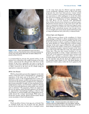
Figure 11.44. Brass band attached to dorsal hoof wall to (white line). This area may remain localized or it may
stabilize a toe crack. Note that the contour of the band matches the progress proximally to form a larger separation in the
coronet. Also note that the defect does not extend distally to the hoof wall. As the separation becomes more extensive and
ground surface of the foot.
extends into a quarter, a concavity (dish) can be seen
forming along one side of the hoof, and a bulge is present
is recommended to increase the ground surface in the on the opposite side directly above the affected area at
palmar foot, redistribute the weight‐bearing on the solar the coronary band (Figure 11.45). The distal phalanx is
surface, and unload the affected heel. The horn in this stabilized or held in place within the hoof capsule by the
section of the foot is generally too thin to accept an
implant, and therefore the hoof wall palmar to the defect
is either removed or the heel can be rebuilt using an
appropriate composite material.
White Line Disease
WLD is a keratolytic process that originates on the solar
surface of the hoof and is characterized by a progressive
separation of the inner zone of the hoof wall. 15,17,21 The
separation occurs in the nonpigmented horn at the junc
tion between the stratum medium and stratum internum. A
separation in the hoof wall is considered to be a delaminat
ing process potentially brought on from mechanical stress,
inappropriate farriery, environmental conditions affecting
the inner hoof wall attachment, and possibly some toxicity
such as selenium. The separation, which can originate at
15
the toe, the quarter, and/or the heel, appears to be invaded
by opportunistic bacteria/fungi leading to infection that
can progress to varying heights and configurations proxi
mally toward the coronet. Interestingly, WLD does not
invade the coronet, and the disease process nearly always
occurs secondary to hoof wall separation.
Etiology
Figure 11.45. Extensive separation under lateral hoof wall
WLD can affect a horse of any age, sex, or breed. One causing a shift of the distal phalanx in the hoof capsule. Note the
or multiple hoofs may be involved and the affected concavity in the medial wall, bulging of the lateral wall, and the soft
hooves can be barefoot or shod. One or multiple horses tissue enlargement above the coronet on the lateral side.