
Page 1158 - Adams and Stashak's Lameness in Horses, 7th Edition
P. 1158
1124 Chapter 11
lamellae; therefore, any significant loss of epidermal
lamellae on the side with the separation allows the distal
VetBooks.ir Other clinical signs may be evident including slow hoof
phalanx to shift toward the side with the separation.
wall growth dorsal to the separation, poor consistency of
hoof wall, and a hollow sound noted when the outer
hoof wall over the separation is tapped with a ham
mer. 17,21 Unfortunately, the disease often goes undetected
until the horse begins to show discomfort.
Radiography can be very informative and should be
considered necessary if WLD is advanced and causing
lameness. Good‐quality radiographs can show the extent
of the hoof wall separation and whether rotation of the
distal phalanx within the hoof capsule has occurred.
Radiography is helpful to differentiate between WLD
and laminitis and can also be used as a guide when
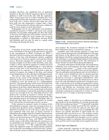
trimming and shoeing the horse. Figure 11.46. Dorsal hoof wall resection. Note the solid margins
around the periphery of the resection.
Farriery
and continue the treatment schedule for WLD as the
Correction of any hoof capsule distortion that may hoof wall grows down is essential for success.
have contributed to the hoof wall separation is essential Complete hoof wall resection (removal of outer hoof
in the management of WLD. Treatment for WLD is wall to expose diseased area) and debridement of all tracts
directed toward protecting and unloading the damaged and fissures in the affected area are often necessary. The
section of the foot with therapeutic farriery combined debridement should be continued proximally and margin
with resection of the hoof capsule overlying the affected ally until there is a solid attachment between the hoof wall
area(s). Resection disrupts the continuity and weight‐ and external lamellae (Figure 11.46). The veterinarian or
bearing strength of the hoof wall; therefore, some type farrier should not reach blood during debridement.
of shoe or device should be applied to stabilize the hoof Treatment with topical disinfectants and medications
wall and transfer the weight‐bearing to a non‐affected following hoof wall resection has been described and
section of the foot. If the separated area of the foot is remains controversial. 17,21,37 None of the topical prepa
determined to be extensive, it is important to plan the rations being used to treat WLD have any proven efficacy.
method of support, the design of the shoe, and method Various antiseptics and astringents such as merthiolate or
of attachment prior to the outer hoof wall being resected. 2% iodine may be helpful not only as a drying agent but
The type of shoe used and the method of support as a dye marker to outline the remaining tracts that
depend on the extent of the damaged hoof wall to be should be removed in subsequent debridement. After
removed. As the toe is involved in most cases of WLD, it thorough hoof wall resection, the affected area is left to
is helpful to move the break‐over in a palmar/plantar grow out with debridement performed at frequent inter
direction. The ground surface of the foot is trimmed from vals. A wire brush can be an effective method for owners
the apex of the frog palmarly, thus creating two planes on to keep the resected area clean daily. The clinician should
the bottom of the foot, which effectively unloads the toe. explore and debride any remaining tracts at 2‐week
The shoe is fitted, so break‐over is just dorsal to the distal intervals. When all tracts are resolved and grown out, a
phalanx in an attempt to unload the dorsal hoof wall and continued examination is indicated at routine re‐shoeing
remove any leverage at the toe. Unloading the hoof wall intervals every 4–5 weeks to prevent recurrence.
distal to the resection is essential to remove the stresses
from the inner hoof wall and promote good growth. This Equine Canker
also minimizes pain by preventing the “pinching” effect at
break‐over that often occurs at the junction of normal Equine canker is described as an infectious process
hoof wall and the proximal aspect of the resection. that results in the development of a chronic hypertrophy
If the resection is extensive and/or if rotation of the of the horn‐producing tissues. 16,27,41 It generally originates
distal phalanx is present, some type of support shoe or a in the frog and may remain focal but has the capacity to
wooden shoe should be used to stabilize the foot. An become diffuse and invade the adjacent sole, bars, and
25
alternative method is to use a bar shoe or open shoe and hoof wall. Canker can occur in one or multiple feet. The
pad combined with some type of silastic material to disease was initially described in draft breeds but can
redistribute the weight on the foot (Equilox International, commonly affect any breed or sex. The etiology remains
Inc., Pine Island, MN). If necessary, attaching glue‐on elusive, but wet environmental or moist, unhygienic con
shoes to the ground surface of the foot is another useful ditions have traditionally been thought to act as a stimu
option for shoeing the horse with WLD. However, lus. However, canker is commonly seen in horses that are
28
acrylic repair of the defect following the resection is not well cared for and in horses that receive regular hoof care.
recommended and should only be considered after all
tracts are resolved. 15,17,21,37 The composite hides and/or Clinical Signs and Diagnosis
fosters the organisms under the repair, and the composite
may weaken the surrounding normal hoof wall. Commit Canker generally originates in the frog and can be
ment from the owner to institute environmental changes mistaken for thrush in the early stages. Thrush is limited