
Page 1163 - Adams and Stashak's Lameness in Horses, 7th Edition
P. 1163
Foot Care and Farriery 1129
VetBooks.ir
Figure 11.51. Lateral radiograph of a horse with thin soles that
would be prone to sole bruising.
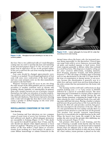
Figure 11.50. Fiberglass foot cast extending to the level of the
proximal pastern.
dermal tissue above the horny sole, the increased pres
sure being responsible for the discomfort. Clinical signs
the foot. One to two additional rolls of 3‐inch fiberglass can range from mild to severe lameness, increased digi
casting tape are used to envelop the foot and extending tal pulse, and marked response to hoof testers. Hoof
to the proximal pastern (Figure 11.50). An acrylic com tester response generally is diffuse rather than isolated
posite may be applied to the toe on the ground surface to a small focal area. The diagnosis is made by ruling out
of the cast to prevent the cast from wearing through the the other common differentials, including laminitis,
ground surface. hoof abscesses, puncture wounds, and distal phalanx
Foot casts should be changed approximately every fractures. 10,11 The old adage of finding signs of bruising
3 weeks or as needed. The prolonged application of foot such as red discoloration in the sole of a lame horse at
casts will cause some contraction of the hoof capsule the time of examination or during farriery is not accu
and may cause pressure sores on the skin immediately rate, because this discoloration is generally seen in a
proximal to the heel bulbs. white foot and may result from previous hemorrhage in
Marginal fractures (type VI) occur at any site along the solar corium that has permeated into the horny sole
the solar margin of the distal phalanx and are commonly over time.
secondary to another condition such as chronic sole The bruising resolves with stall confinement on deep
bruising, chronic laminitis, or osteomyelitis. In general, sawdust bedding and a 3‐ to 5‐day course of NSAIDs
these solar margin fractures are not of primary concern followed by a period of controlled exercise. The most
themselves, but rather an indication of another underly important part of treatment is a farriery plan to improve
ing condition such as foot with limited protection due to sole depth. Farriery options include leaving the sole
very thin sole thickness. These horses respond to shoeing intact during trimming, evaluating the length of the toe
with a wide‐web straight‐bar shoe that is well concaved when placing the shoe, modifying break‐over, and add
(using a grinder or skilled forging) on the inner solar ing some mild heel elevation. Placing a leather or plastic
surface of the shoe. pad between the ground surface of the foot and the shoe
protects the thin sole and provides comfort, but gener
ally does not improve the structures of the foot. The use
MISCELLANEOUS CONDITIONS OF THE FOOT of a rim pad between the foot surface and the shoe is a
Sole Bruising common farrier practice to raise the foot off the ground.
This practice may provide transient improvement;
Foot bruising and foot infections are two common however, it concentrates the weight‐bearing on the wall.
causes of acute onset of severe foot lameness. Feet that When the horse’s foot loads, the weight of the horse
lack sufficient sole depth to afford adequate protection pushes the sole toward the ground, thus inhibiting any
are susceptible to bruising (Figure 11.51). Sole depth further sole growth (Figure 11.52).
44
for a good foot should be greater than 15 mm, and the Sole pressure often leads to sole bruising, and this
sole should not deform when digital pressure or hoof type of bruising is usually noted shortly after the horse
testers are applied. Bruising can occur when the ground is trimmed or shod. In this case the hoof wall is trimmed
39
surface of the foot strikes a hard object or excessive shorter than the adjacent sole so that when a shoe is
trauma from working on a hard surface. It appears to applied to the foot and clinched or when the horse stands
cause diffuse hemorrhage or edema formation in the on a hard surface, excess pressure is placed on the sole.