Page 312 - Adams and Stashak's Lameness in Horses, 7th Edition
P. 312

VetBooks.ir


                                                                                                               1
                                                           10
                                                                                                               2


                                                                                                              3
                                                            12
                                                            11
                                                                                                             4
                                                                                                             5
                                                                5
                                                                                                            6
                                                                                                            2
                                                                  10                                       1

                                                                                                         7
                                                                                                      8


                                                                                             9
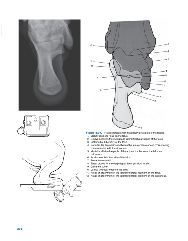
                                                         Figure 3.77.  Flexed dorsoplantar (flexed DP) projection of the tarsus.
                                                          1.  Medial trochlear ridge on the talus.
                                                          2.  Groove between the medial and lateral trochlear ridges of the talus.
                                                          3.  Distomedial tuberosity of the talus.
                                                          4.  Nonarticular depressions between the talus and calcaneus. This opening
                                                            communicates with the sinus tarsi.
                                                          5.  Medial and lateral aspects of the articulation between the talus and
                                                            calcaneus.
                                                          6.  Proximomedial tuberosity of the talus.
                                                          7.  Sustentaculum tali.
                                                          8.  Tarsal groove for the deep digital flexor principal tendon.
                                                          9.  Calcaneal tuber.
                                                         10.  Lateral trochlear ridge on the talus.
                                                         11.  Areas of attachment of the lateral collateral ligament on the talus.
                                                         12.  Areas of attachment of the lateral collateral ligament on the calcaneus.


            278
   307   308   309   310   311   312   313   314   315   316   317