
Page 317 - Adams and Stashak's Lameness in Horses, 7th Edition
P. 317
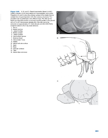
Figure 3.80.  (A, B, and C). Flexed lateromedial (flexed L0‐20Cr‐  A
             CaMO) projection of the femorotibial and femoropatellar joints (stifle).
  VetBooks.ir  condyle free of superimposition with other osseous structures. This
             Projections (A and C) show the articular surface of the medial femoral
             projection can be performed in two different ways. The limb can be
             flexed and abducted and the X‐ray beam directed parallel to the ground
             with a 0° to 20° laterocranial obliquity (B); if the limb cannot be
             abducted, an approximately 10° distal obliquity (L0‐20Cr10Di‐PrMO)
             should be added to the X‐ray beam direction.
              1.  Femur
              2.  Medial trochlea
              3.  Lateral trochlea
              4.  Medial condyle
              5.  Lateral condyle
              6.  Intertrochlear groove
              7.  Extensor fossa                                  B
              8.  Intercondylar fossa
              9.  Patella
             10.  Medial articular surface
             11.  Apex
             12.  Base
             13.  Articular surfaces
             14.  Tibia
             15.  Medial tibial eminence


                                                                  C


                                                                                                             283