
Page 314 - Adams and Stashak's Lameness in Horses, 7th Edition
P. 314
VetBooks.ir
31
26
a 1
30
29 b
28
27 2
3
26
25 4
24
23
22 5
21 6
20 7
8
19
18 9
17 10
16 11
12
15
c 13
14
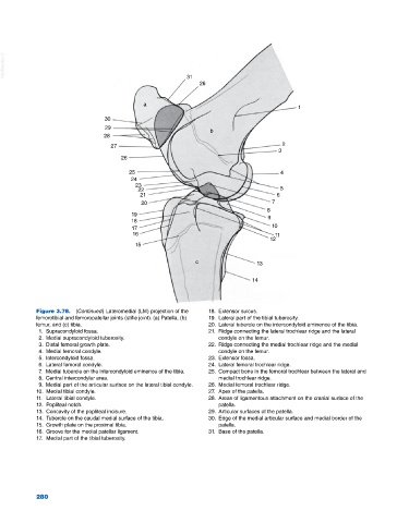
Figure 3.78. (Continued) Lateromedial (LM) projection of the 18. Extensor sulcus.
femorotibial and femoropatellar joints (stifle joint). (a) Patella, (b) 19. Lateral part of the tibial tuberosity.
femur, and (c) tibia. 20. Lateral tubercle on the intercondyloid eminence of the tibia.
1. Supracondyloid fossa. 21. Ridge connecting the lateral trochlear ridge and the lateral
2. Medial supracondyloid tuberosity. condyle on the femur.
3. Distal femoral growth plate. 22. Ridge connecting the medial trochlear ridge and the medial
4. Medial femoral condyle. condyle on the femur.
5. Intercondyloid fossa. 23. Extensor fossa.
6. Lateral femoral condyle. 24. Lateral femoral trochlear ridge.
7. Medial tubercle on the intercondyloid eminence of the tibia. 25. Compact bone in the femoral trochlear between the lateral and
8. Central intercondylar area. medial trochlear ridge.
9. Medial part of the articular surface on the lateral tibial condyle. 26. Medial femoral trochlear ridge.
10. Medial tibial condyle. 27. Apex of the patella.
11. Lateral tibial condyle. 28. Areas of ligamentous attachment on the cranial surface of the
12. Popliteal notch. patella.
13. Concavity of the popliteal incisure. 29. Articular surfaces of the patella.
14. Tubercle on the caudal medial surface of the tibia. 30. Edge of the medial articular surface and medial border of the
15. Growth plate on the proximal tibia. patella.
16. Groove for the medial patellar ligament. 31. Base of the patella.
17. Medial part of the tibial tuberosity.
280