
Page 436 - Adams and Stashak's Lameness in Horses, 7th Edition
P. 436
402 Chapter 3
VetBooks.ir
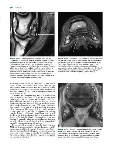
Figure 3.225. Sagittal proton density image of the foot of a Figure 3.226. Transverse T2* gradient echo image of the foot of
horse with chronic navicular bone degeneration. There is localized a horse with chronic navicular bone disease. The palmar border of
signal hyperintensity in the distal third of the flexor border of the the navicular bone is irregular due to degenerative erosion of the
navicular bone due to loss of cortical bone and the presence of flexor cortex. There is sclerosis of the medullary cavity of the
fibrous tissue and bursal synovial fluid in the defect (arrow). There is navicular bone. The normal hyperintense synovial fluid layer
diffuse loss of signal in the medullary cavity of the navicular bone separating the palmar border of the navicular bone from the dorsal
due to osteosclerosis. There is loss of separation between the surface of the deep digital flexor tendon is interrupted by hypoin
palmar surfaces of the thickened collateral sesamoidean and distal tense fibrous adhesions between both surfaces (arrows).
sesamoidean impar ligaments on the one hand and the dorsal
surface of the deep digital flexor tendon on the other, suggestive of
abnormal adherence between these structures.
frequently accompanied by fibrillation of the dorsal
aspect of the DDFT. Areas of advanced fibrocartilage
and cortical bone loss from the flexor surface of the
navicular bone are prone to adhesion formation between
the dorsal surface of the DDFT and the exposed area of
bone (Figure 3.226).
Another form of degenerative navicular bone disease
may be caused by the presence of osseous fragments at the
distal border of the navicular bone (Figure 3.227). 8–10,50
Especially large fragments may induce a focal remodeling
response in the distal margin of the navicular bone, result
ing in localized osseous fluid with STIR signal hyperinten
sity, irregular thickening of cortical bone, and formation
of an osseous cyst‐like lesion. A distal border fragment
9
accompanied by focal medullary bone edema, an osseous
cyst‐like lesion, or thickened cortical bone at the distal
border of the navicular bone is likely to be associated with
lameness. Even so, distal border fragments may be asymp
tomatic, especially when they are smaller and if there is
198
no MRI evidence of remodeling of the distal border of the
navicular bone adjacent to the fragment.
In horses with a complete navicular bone fracture,
MRI can demonstrate the fracture configuration on 3D
images and elucidate if fracture fragments have lacer Figure 3.227. Dorsal T1‐weighted fast low‐angle shot (FLASH)
ated the surface of the DDFT, prior to possible attempts image at the level of the navicular bone, oriented parallel with its
at surgical repair. Fractures must be distinguished from flexor surface. The narrow slice thickness and orientation of this
bipartite navicular bones for the purpose of prognosis sequence allow for identification of an osseous fragment at the
and treatment planning. 76 lateral angle of the distal border of the navicular bone (arrow).