
Page 437 - Adams and Stashak's Lameness in Horses, 7th Edition
P. 437
Diagnostic Imaging 403
Lesions of the Distal Sesamoidean Impar Ligament lesion (Figure 3.228). Generalized thickening of the
199
impar ligament and adhesions between the impar liga
The distal sesamoidean impar ligament is outlined by
VetBooks.ir hyperintense fluid signal from the DIP joint dorsally and ment and the DDFT occur most commonly in associa
tion with navicular bursitis accompanying marked
the distal recess of the navicular bursa distally. It is com
pathology of the DDFT or the navicular bone and may
posed of individual fiber bundles with synovial inter
digitations of the DIP joint and small branches of the be an integral part of secondary generalized bursal
inflammation.
palmar digital arteries. The heterogeneous structure
with multiple fluid–fiber interfaces in this small liga
ment results in a high susceptibility to partial volume Lesions of the Collateral Sesamoidean Ligaments
averaging. Therefore, focal high signal within the impar It has been reported that the paired collateral sesa
ligament must be interpreted with caution because of
normal signal variation. One study found no significant moidean ligaments have uniform low intensity signal
in all image sequences and are symmetrical in thickness
correlation between the MRI appearance of the impar 152
ligament itself and the presence of histological abnor medially and laterally. However, the presence of sig
nal increase is a common normal variation on all con
malities. Moreover, the ligament is thicker and its pal
50
mar border more intimately apposed to the dorsal trast weightings, especially near the insertion to the
The dorsal
proximal border of the navicular bone.
17,176
border of the DDFT axially than abaxially, which may
confound identification of ligament thickening and and palmar borders of the ligaments are clearly demar
cated by high fluid signal in the palmar recess of the
adhesion formation. A diagnosis of desmitis of this liga
ment is rare and rarely considered the primary cause of DIP joint and the proximal recess of the navicular
bursa.
lameness, except in the presence of distal border frag
Collateral sesamoidean ligament injury may be evi
ments or cystic lesions in the distal border of the navicu dent as an altered shape with focal or diffuse signal
lar bone. 50
Unequivocal MRI signs of impar ligament injury increase in the body of the ligament. Asymmetric thick
include marked thickening, extensive adhesion of the ening is most easily identified when the paired ligament
palmar surface of the ligament to the dorsal surface of is normal (Figure 3.229). Generalized thickening of the
ligament with loss of separation from the dorsal surface
the DDFT with loss of normal fluid space in the distal
recess of the navicular bursa, and osseous signal abnor of the DDFT, possibly with adhesions, occurs most com
monly in association with chronic navicular bursitis sec
malities at the insertion of the ligament to the distal pha
lanx. The latter include focally increased fluid signal, ondary to tendinopathy or degenerative navicular bone
disease.
increased mineralization, entheseous new bone produc
tion, and focal osteolysis with formation of a cyst‐like Primary collateral sesamoidean ligament injury is
rare except in one report of horses with hindfoot
lameness. 11
Lesions of the Navicular Bursa
Pooling of hyperintense fluid can normally be
observed in the proximolateral and proximomedial
pouches of the navicular bursa and to a lesser extent
between the DDFT and the distal impar ligament dis
tally. There should be visible separation between the
dorsal surface of the DDFT and the palmar surfaces of
the collateral and distal impar sesamoidean ligaments in
normal navicular bursae, apart from the areas of attach
ment between these structures by the normal proximal
and distal synovial membrane reflections in the sagittal
midline of the navicular bursa.
Navicular bursitis results in effusion with enlarge
ment of the proximolateral and proximomedial
pouches of the bursa and sometimes in dorsal devia
tion of the central part of the collateral sesamoidean
ligaments or the distal impar ligament. The proximo
lateral recess of an inflamed navicular bursa is always
more distended than the proximomedial recess. Fibrous
scar tissue can occur in the proximal and distal recesses
of the bursa and is indicative of chronic bursitis. Lack
of separation between the dorsal surface of the DDFT
and the palmar surface of the collateral sesamoidean
79
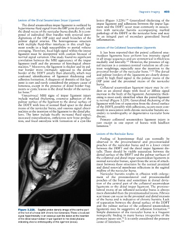
Figure 3.228. Sagittal proton density image of the central part ligaments may be suggestive of adhesion formation.
of the foot of a horse with chronic foot lameness. There is localized Simple distension of the navicular bursa is a frequent
signal hyperintensity in an osseous cyst‐like lesion at the insertion nonspecific finding in many horses irrespective of the
46
of the distal sesamoidean impar ligament to the distal phalanx primary injury site. It is rarely considered the primary
indicating chronic enthesopathy of this ligament (arrow). cause of lameness. 152