
Page 433 - Adams and Stashak's Lameness in Horses, 7th Edition
P. 433
VetBooks.ir
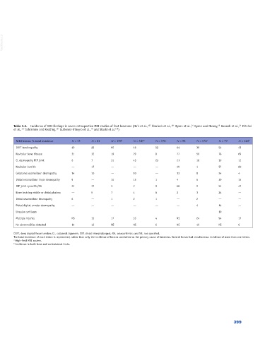
Table 3.3. Incidence of MRI findings in seven retrospective MRI studies of foot lameness (Mair et al., Sherlock et al., Dyson et al., Dyson and Murray, Boswell et al., Mitchel
54
164
107
24
49
et al., Schramme and Redding, Gutierrez‐Nibeyro et al., and Stockl et al. )
76
179
157
113
MRI lesions % total incidence N = 35 N = 41 N = 199 a N = 347 a N = 170 N = 98 N = 172 a N = 79 N = 120 b
DDFT tendinopathy 49 29 60 48 52 64 30 54 43
Navicular bone disease 31 32 19 29 8 77 50 78 69
CL desmopathy DIP joint 6 7 31 43 23 21 16 39 12
Navicular bursitis — 17 — — — 49 1 57 60
Collateral sesamoidean desmopathy 14 10 — 10 — 13 8 24 4
Distal sesamoidean impar desmopathy 9 — 10 15 1 4 6 30 15
DIP joint synovitis/OA 23 37 3 2 9 68 9 53 47
Bone bruising middle or distal phalanx — 5 7 4 6 2 3 24 —
Distal sesamoidean desmopathy 6 — 1 2 1 — 2 — —
Distal digital annular desmopathy — — — — — — 4 14 —
Ungular cartilages 10
Multiple injuries NS 15 17 33 4 NS 24 94 17
No abnormalities detected 14 12 NS NS 6 NS 10 NS 6
DDFT, deep digital flexor tendon; CL, collateral ligament; DIP, distal interphalangeal; OA, osteoarthritis; and NS, not specified.
The total incidence of each lesion is represented, rather than only the incidence of lesions considered as the primary cause of lameness. Several horses had simultaneous incidence of more than one lesion.
a High‐field MRI system.
b Incidence in both lame and contralateral limbs.
399