
Page 490 - Adams and Stashak's Lameness in Horses, 7th Edition
P. 490
456 Chapter 4
callus. Computer‐assisted surgery systems may provide use, complete radiographic healing did not occur in any
81
123
further advantages in screw placement in the future. 54 case in this study. Important clinical aspects of navicu-
VetBooks.ir formed to relieve pain in cases that have not responded clinical studies include the following:
Palmar or plantar digital neurectomy can also be per-
lar bone fractures that can be summarized from these
to conservative treatment.
The navicular bone is very
5,72
slow to heal, and these fractures are invariably associ- 1. Heel elevation should be an important aspect of
treatment.
ated with damage to the impar ligament, DDFT, and
DIP joint. 5,60 Chronic lameness may result from poor 2. Four to 6 months of confinement may be necessary.
3. Complete fracture healing is unlikely to occur.
fracture healing and adhesions that develop between the
72
DDFT and the navicular bone. Follow‐up radiography 4. A PD neurectomy will minimize the lameness but
does not guarantee a sound horse.
on 17 horses with complete navicular bone fractures
revealed increases in the width of the fracture gap for up
to 4 months after injury. A noncalcified fibrous union
72
can still be evident years after the fracture occurred, SOFT TISSUE INJURIES IN THE FOOT (DDFT
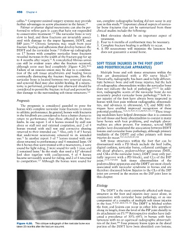
although some may heal completely (Figure 4.20). 4,116 AND PODOTROCHLEAR APPARATUS)
Failure of an osseous union may be due to a combina-
tion of the soft tissue attachments and loading forces Multiple bone and soft tissue structures within the
111
continually distracting the fracture fragments. Also the foot are desensitized with a PD nerve block.
navicular bone is located between two synovial spaces, Historically, radiography has been used to help differen-
and synovial fluid may also inhibit healing. If a neurec- tiate between bone and soft tissue injuries, but the lack
tomy is performed, prolonged rest afterward should be of radiographic abnormalities within the navicular bone
considered to permit the fracture to heal and prevent fur- does not indicate the lack of pathology. 2,84,101 In addi-
ther damage to the surrounding soft tissue structures. 116 tion, radiographic scores of the navicular bone do not
accurately predict navicular bone pathology. Soft tis-
84
sue injuries of the foot have always been suspected in
Prognosis horses with foot pain without radiographic abnormali-
The prognosis is considered guarded to poor for ties, and advances in ultrasound, CT, and MRI tech-
horses with complete navicular bone fractures to return niques have enabled improved recognition of these
to athletic performance. In general, horses with fractures potential problems. 19,20,38,39,41–45,56,84,90,91,125,131 These imag-
in the hindlimb are considered to have a better chance to ing modalities have helped determine that it is common
return to performance than those affected in the fore- for soft tissue and bony abnormalities to coexist in many
limbs. In one report 3 of 6 horses treated by stall rest lame horses with foot problems, and multiple abnor-
alone or stall rest and external coaptation, and 2 of 4 malities are often present. For instance, several MRI
horses treated with stall rest and corrective shoeing studies have documented the association between DDFT
returned to their intended use. Also, only 1 of 5 horses lesions and navicular bone pathology, although primary
72
that underwent neurectomy returned to its intended tendinitis of the DDFT and other primary soft tissue
use. In another report 3 of 7 horses treated with rest injuries do occur. 39,41,42,44,84,105,112
72
alone returned to performance and 4 remained lame. Of Soft tissue structures within the foot that can be
the 6 horses that were treated with a neurectomy, 2 were desensitized with a PD block include the heel bulbs,
sound for light riding, 2 were sound for only 1 year, and digital cushion, navicular bursa, collateral cartilages of
2 remained lame. In the study that used a 12° elevated the distal phalanx, podotrochlear apparatus (DSIL,
5
heel shoe together with confinement, 4 of 4 horses and CSLs of the navicular bone), DDFT (may only par-
became serviceably sound for riding, and 2 of 4 returned tially improve with a PD block), and CLs of the DIP
123
to competition. Although the horses were sound for joint. 35,42,44,84,111 Soft tissue abnormalities of the
podotrochlear apparatus and the DDFT are most closely
associated with navicular syndrome in horses and are
therefore discussed below. Injuries to the CLs of the DIP
joint are covered in the section on the DIP joint later in
this chapter.
Etiology
The DDFT is the most commonly affected soft tissue
structure in the foot and injuries may occur alone, in
conjunction with navicular bone pathology, or as one
component of a complex of multiple soft tissue injuries
in the foot. 38,39,41–44,84,104–106 The DDFT is bilobed within
the foot, and lesions can occur in either lobe anywhere
along its length, from the level of the PIP joint distally to
104
its attachment on P3. Retrospective studies have indi-
cated a prevalence of 30%–64% in horses with foot
lameness with no or equivocal radiographic abnormali-
106
Figure 4.20. This oblique radiograph of the navicular bone was ties of the foot. Four primary lesion types in the distal
taken 23 months after the fracture occurred. portion of the DDFT have been identified: core lesions,