
Page 492 - Adams and Stashak's Lameness in Horses, 7th Edition
P. 492
458 Chapter 4
pathology. However, there is often a history of an acute
onset of moderate to severe lameness that may improve
VetBooks.ir history of activity that caused excessive hyperextension
There may be a
with rest and worsen with exercise.
35,36
of the foot such as working in soft ground and/or jump-
ing. The lameness may be unilateral and may worsen in
a circle or when exercised on soft ground. Occasionally,
pain may be elicited with deep palpation of the DDFT
between the collateral cartilages of the heels. Hoof tester
pain is variable but may be present if a DDFT lesion and
navicular pathology are present concurrently. Phalangeal
flexion may cause a positive response but is often vari-
able. Increasing tension on the DDFT with the navicular
wedge test may accentuate the lameness. 35,36
The lameness is not reliably abolished with a PD
nerve block in horses with DDFT lesions. 36,42 Horses
often improve but in one study only 24% of those with
DDF tendinitis responded completely to a PD nerve
block. The lameness should respond to basisesamoid
42
block and many improve after IA anesthesia of the DIP
joint. Anesthesia of the DIP joint was more effective in
alleviating the lameness in horses with DDFT lesions
than was the PD block in one study. This finding dif-
42
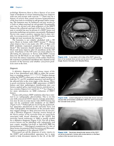
fers from the clinical experience of the author. However, Figure 4.22. A core lesion within lobe of the DDFT above the
the response to perineural anesthesia may depend on the level of the navicular bone (arrow) can be seen in this PD axial MR
location of the lesion(s) and whether concurrent prob- image. Source: Courtesy of Dr. Natasha Werpy.
lems exist in the foot.
Diagnosis
A definitive diagnosis of a soft tissue injury of the
foot is best determined with MRI in either the recum-
bent or standing patient. 38,39,41–44,78,105,106 Tendon damage
within the DDFT is often seen as focal signal increase on
both the T1‐ and T2‐weighted sequences and swelling of
the affected lobe in the acute stages of the disease. There
is good correlation between MRI appearance of DDFT
lesions and their pathological classification into core
lesions, sagittal splits, insertional lesions, and dorsal sur-
face erosions (Figures 4.21 and 4.22). 104,106 See Chapter 4
for more information on MRI evaluation of DDFT
lesions.
Horses with primary DDF tendinitis often have mini-
mal to no radiographic abnormalities. This is not the
case with concurrent DDFT injuries. Ectopic mineraliza- Figure 4.23. Lateral radiograph of a horse with chronic navicular
tion may be seen in some horses with DDFT lesions but disease that demonstrates calcification within the DDFT proximal to
the navicular bone (arrow).
may not be correlated with tendinitis of the DDFT
(Figure 4.23). Enthesophytes involving the podotroch-
36
lear apparatus attachments to the navicular bone and
other radiographic abnormalities of the navicular bone
may suggest damage to these structures but do not pro-
vide a definitive diagnosis. Likewise, erosive lesions of
the flexor surface of the navicular bone are most likely
associated with dorsal abrasions of the DDFT, but
navicular bursal endoscopy or MRI is usually needed
for a definitive diagnosis (Figure 4.24). Not all of these
erosive lesions can be identified with radiography and
may require standing low‐field or high‐field MRI for
documentation. Histological examination of a few of
112
these horses documented fibril formation and fibrocarti-
laginous metaplasia of the adjacent DDFT. 112
Ultrasound can aid the diagnosis of some injuries to Figure 4.24. Secondary dorsal border lesions of the DDFT
the DDFT and podotrochlear apparatus but is most should be suspected in horses with erosive lesions on the flexor
useful to help document injuries to the CL of the DIP surface of the navicular bone (arrow).