
Page 678 - Fluid, Electrolyte, and Acid-Base Disorders in Small Animal Practice
P. 678
CHAPTER • 28
Peritoneal Dialysis
Linda A. Ross and Mary Anna Labato
Dialysis is the process by which water and solutes move Thesevesselsarelocatedatvariousdistancesfromthemeso-
between two compartments that are separated by a semi- thelial surface and can be found throughout the connective
permeable membrane. In peritoneal dialysis (PD), the tissue layer. Lymphatics also are found in this layer, most
two compartments consist of blood in the peritoneal commonly in the subdiaphragmatic peritoneum. These
capillaries and fluid (dialysate) instilled into the peritoneal lymphatics drain primarily via stomata in the diaphragmatic
cavity; the peritoneum serves as the semipermeable peritoneum. 10,48 The role of lymphatics in fluid and solute
membrane. The primary indication for PD in animals is exchange from the peritoneum is poorly understood
for renal failure to correct the resulting water, solute, because of the difficulty in directly measuring lymph flow.
and acid-base abnormalities and to remove uremic toxins. Lymph flow is affected more by gravity than is blood flow
through vessels, and therefore the upright posture of
BIOLOGY OF THE humans versus the quadruped stance of animals may mean
PERITONEAL MEMBRANE that the role of peritoneal lymphatics differs between
species.
The peritoneum is the serosal membrane that lines the The most important function of the peritoneal mem-
abdominal cavity. The portion that covers the viscera and brane is to provide a protective, lubricating surface for the
other intraabdominal structures is known as the visceral abdominal organs. Mesothelial cells secrete glycosami-
peritoneum, and that which lines the abdominal cavity is noglycans including hyaluronic acid, proteoglycans such
known as the parietal peritoneum. In humans, the surface
areaoftheperitoneumisapproximatelythesameasthebody
2
surface area (1 to 2 m ), and the visceral peritoneum
accounts for approximately 80% of the total. 10 Peritoneal
surface area isproportionately largerincomparisonto body
surface area in infants and children, 11 suggesting that this
difference would also be true for dogs and cats.
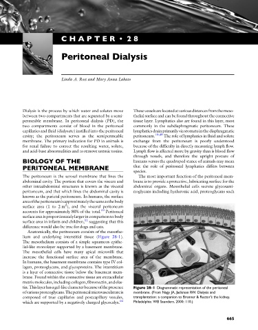
Anatomically, the peritoneum consists of the mesothe-
lium and underlying interstitial tissue (Figure 28-1).
The mesothelium consists of a simple squamous epithe-
lial-like monolayer supported by a basement membrane.
The mesothelial cells have many apical microvilli that
increase the functional surface area of the membrane.
In humans, the basement membrane contains type IV col-
lagen, proteoglycans, and glycoproteins. The interstitium
is a layer of connective tissue below the basement mem-
brane. Found within the connective tissue are extracellular
matrix molecules, including collagen, fibronectin, and elas-
tin.Thislayerhasagel-likecharacterbecauseofthepresence Figure 28-1 Diagrammatic representation of the peritoneal
ofvariousproteoglycans.Theperitonealmicrovasculatureis membrane. (From Nagy JA, Jackman RW. Dialysis and
composed of true capillaries and postcapillary venules, transplantation: a companion to Brenner & Rector's the kidney.
which are supported by a negatively charged glycocalyx. 61 Philadelphia: WB Saunders, 2000: 110.)
665