
Page 208 - Basic Monitoring in Canine and Feline Emergency Patients
P. 208
exercise, or defecation, so small variations are
expected to occur. The IAP may also vary with level
VetBooks.ir Manometer of sedation, body positioning, body condition
column
score, and bladder instillation volumes during IAP
testing (see below). As much as possible, IAP
should be obtained the same way every time to
minimize variations in the results.
The presence of IAH has been reported in veter-
inary patients. In one study evaluating intra-
abdominal pressures in 20 client dogs after routine
ovariohysterectomy, there were significant eleva-
tions in the mean postoperative pressure by as
much as 15 cmH O (Conzemius et al., 1995). This
2
increase in IAP persisted for as long as 24 hours
Line to Extension after the elective surgery. However, there was no
patient to fill clinical evidence of significant complications in the
Manometer
group of healthy dogs. Intra-abdominal pressures
were also measured in 20 dogs with gross abdom-
inal distension (due to clinical conditions like gas-
tric dilation and volvulus, closed pyometra,
hemoperitoneum, and ascites). All of these dogs
had severe IAP (>16 cmH O) either before or after
2
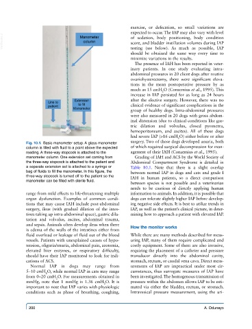
Fig. 10.1. Basic manometer setup. A glass manometer surgery. Two of those dogs developed anuria, both
column is filled with fluid to a point above the expected of which required surgical decompression for man-
reading. A three-way stopcock is attached to the agement of their IAH (Conzemius et al., 1995).
manometer column. One extension set coming from Grading of IAH and ACS by the World Society of
the three-way stopcock is attached to the patient and Abdominal Compartment Syndrome is detailed in
a separate extension set is attached to a syringe or Table 10.1. Note that there is a slight overlap
bag of fluids to fill the manometer. In this figure, the between normal IAP in dogs and cats and grade I
three-way stopcock is turned off to the patient so the IAH in human patients, so a direct comparison
manometer can be filled with sterile fluid.
between species is not possible and a veterinarian
needs to be cautious of directly applying human
range from mild effects to life-threatening multiple information to animals. In addition, it is possible that
organ dysfunction. Examples of common condi- dogs can tolerate slightly higher IAP before develop-
tions that may cause IAH include post-abdominal ing negative side effects. It is best to utilize trends in
surgery, ileus (with gradual dilation of the intes- IAP, as well as the patient’s clinical picture, in deter-
tines taking up intra-abdominal space), gastric dila- mining how to approach a patient with elevated IAP.
tation and volvulus, ascites, abdominal trauma,
and sepsis. Animals often develop ileus when there
is edema of the walls of the intestines either from How the monitor works
fluid overload or leakage of fluid out of the blood While there are many methods described for meas-
vessels. Patients with unexplained causes of hypo- uring IAP, many of them require complicated and
tension, oliguria/anuria, abdominal pain, azotemia, costly equipment. Some of them are also invasive,
elevated liver enzymes, or respiratory difficulty, requiring the placement of a catheter and pressure
should have their IAP monitored to look for indi- transducer directly into the abdominal cavity,
cations of ACS. stomach, rectum, or caudal vena cava. Direct meas-
Normal IAP in dogs may range from urements of IAP are impractical under most cir-
5–10 cmH O, while normal IAP in cats may range cumstances, thus surrogate measures of IAP have
2
from 0–20 cmH O. For measurements obtained in been investigated. The homogenous transmission of
2
mmHg, note that 1 mmHg is 1.36 cmH O. It is pressure within the abdomen allows IAP to be esti-
2
important to note that IAP varies with physiologic mated via either the bladder, rectum, or stomach.
conditions such as phase of breathing, coughing, Intravesical pressure measurement, using the uri-
200 A. Odunayo