
Page 212 - Basic Monitoring in Canine and Feline Emergency Patients
P. 212
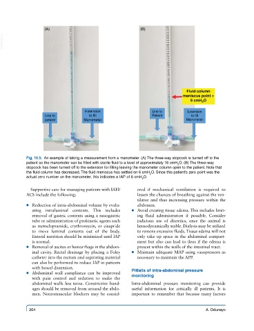
(A)                                     (B)
  VetBooks.ir


                                                                                Fluid column
                                                                              meniscus point =
                                                                                 6 cmH 2 O

                                     Extension                   Line to        Extension
                    Line to            to fill                   Patient          to fill
                    patient         Manometer                                   Manometer


            Fig. 10.5.  An example of taking a measurement from a manometer. (A) The three-way stopcock is turned off to the
            patient so the manometer can be filled with sterile fluid to a level of approximately 16 cmH O. (B) The three-way
                                                                            2
            stopcock has been turned off to the extension for filling leaving the manometer column open to the patient. Note that
            the fluid column has decreased. The fluid meniscus has settled on 6 cmH O. Since this patient’s zero point was the
                                                                2
            actual zero number on the manometer, this indicates a IAP of 6 cmH O.
                                                             2
              Supportive care for managing patients with IAH/  ered if mechanical  ventilation  is required to
            ACS include the following:                     lessen the chances of breathing against the ven-
                                                           tilator and thus increasing pressure within the
             ● ● Reduction of intra-abdominal volume by evalu-  abdomen.
               ating intraluminal contents.  This includes   ● ● Avoid creating tissue edema. This includes limit-
               removal of gastric contents using a nasogastric   ing  fluid  administration  if  possible.  Consider
               tube or administration of prokinetic agents such   judicious use of diuretics, once the animal is
               as metoclopramide, erythromycin, or cisapride   hemodynamically stable. Dialysis may be utilized
               to move luminal contents out of the body.   to remove excessive fluids. Tissue edema will not
               Enteral nutrition should be minimized until IAP   only take up space in the abdominal compart-
               is normal.                                  ment but also can lead to ileus if the edema is
             ● ● Removal of ascites or hemorrhage in the abdom-  present within the walls of the intestinal tract.
               inal cavity. Rectal drainage by placing a Foley   ● ● Maintain adequate MAP using vasopressors as
               catheter into the rectum and aspirating material   necessary to maintain the APP.
               can also be performed to reduce IAP in patients
               with bowel distension.                    Pitfalls of intra-abdominal pressure
             ● ● Abdominal wall compliance can be improved   monitoring
               with pain control and sedation to make the
               abdominal walls less tense. Constrictive band-  Intra-abdominal pressure monitoring can provide
               ages should be removed from around the abdo-  useful information for critically ill patients. It is
               men. Neuromuscular blockers may be consid-  important to remember that because many factors


             204                                                                        A. Odunayo