
Page 211 - Basic Monitoring in Canine and Feline Emergency Patients
P. 211
8. Take the measurement about 30–60 seconds
after instilling saline into the urinary bladder so
VetBooks.ir that the detrusor muscle is relaxed and there is no
active abdominal contraction. The measurement
should ideally be taken at the end of expiration.
9. A clinical decision needs to be made about the
results obtained. If the IAP is high and the patient
has clinical signs of ACS, the clinician should make
the decision to treat the patient. Repeated meas-
urements after treatment may be used to gage
the response to treatment as well as trends in the
patient’s IAP over time.
10. Disconnect three-way stopcock, syringe, saline
bag and extension tubing once finished. Reattach
urine collection bag to Foley catheter in a sterile
manner.
(This method can be performed in a similar manner
using a nasogastric tube in the stomach, in cases
where the urinary bladder cannot be utilized. No
specific technique has been published or validated
in veterinary medicine and the bladder is always
preferred.)
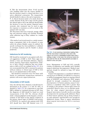
Fig. 10.4. A cat receiving a manometer reading. Note
Indications for IAP monitoring that the manometer is attached to the side of the
cage so as to be in a repeatable location each time a
IAP should be monitored in any patient with clini- reading is taken. In this case, the red arrow marks the
cal suggestions of IAH or ACS. These signs may zero point for this cat. All measurements are taken in
include a tense and distended abdomen, oliguria, relation to this zero point.
anuria, hypoxia, hypercapnia, hypotension, acido-
sis, or ileus. Clinical conditions that may predis- hours. Management of IAH and ACS, typically
pose to IAH or ACS are outlined in Box 10.1. It is consists of supportive care initially, and if needed,
not recommended that IAP is measured in patients surgical abdominal decompression. Medical man-
without evidence or clinical signs of IAH or ACS agement should be tailored to keep IAP below
due to over-interpretation of the results. 15 mmHg.
IAP should be monitored every 4–6 hours with Surgical decompression is considered definitive
hourly (or continuous) measurement restricted to management of IAH/ACS. If IAP/ACS is refractory
patients with severe organ dysfunction. to medical management based on ongoing clinical
signs of IAH/ACS and refractory IAH, surgery
should be strongly considered for abdominal
Interpretation of IAP results
decompression. In human patients, surgical inter-
Once the IAP has been obtained, it should then be vention is considered when IAP is greater than
determined if the patient is at risk for IAH, as 26 mmHg or the patient's clinical signs cannot be
detailed in Table 10.1. It is important to note that controlled. However, there are no absolute guide-
IAH is defined as ‘sustained increases in IAP’, thus lines for when surgical intervention occurs.
the diagnosis of IAH should be based on serial Surgical intervention typically involves providing
measurements of the IAP. The clinician should decompression by opening up the abdomen. Many
make a clinical judgment on the frequency of meas- of these human patients end up being treated as
urement of IAP based on the clinical condition of an ‘open abdomen’ until the IAP is low enough for
the patient, clinical signs of IAH/ACS and severity abdominal closure. There is sparse information
of IAH/ACS. As a general rule, patients with IAH/ about surgical intervention for ACS in veterinary
ACS should have their IAP checked every 2–4 patients.
Manometer-based Monitoring 203