
Page 520 - Feline diagnostic imaging
P. 520
532 30 Peritoneal Cavity
(b)
(a)
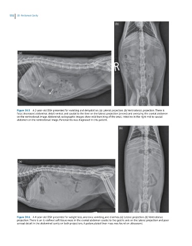
Figure 30.5 A 2-year-old DSH presented for vomiting and dehydration. (a) Lateral projection. (b) Ventrodorsal projection. There is
focal decreased abdominal detail ventral and caudal to the liver on the lateral projection (arrows) and overlying the cranial abdomen
on the ventrodorsal image. Abdominal radiographic images show mild bunching of the small intestine in the right mid to caudal
abdomen on the ventrodorsal image. Pancreatitis was diagnosed in this patient.
(b)
(a)
Figure 30.6 A 4-year-old DSH presented for weight loss, anorexia, vomiting, and diarrhea. (a) Lateral projection. (b) Ventrodorsal
projection. There is an ill-defined soft tissue mass in the cranial abdomen caudal to the gastric axis on the lateral projection and poor
serosal detail in the abdominal cavity on both projections. A pedunculated liver mass was found on ultrasound.