
Page 522 - Feline diagnostic imaging
P. 522
534 30 Peritoneal Cavity
In a more recent study of peritoneal carcinomatosis eval- abdominal organs such as the liver. The liver size can be
uated with CT in humans, the key imaging findings evaluated by locating the stomach axis; a shift of the stom-
included ascites, greater omental invasion with nodules, ach axis caudally and dorsally will signify enlargement of
involvement of the mesentery with masses or nodules, and varying degree. With decreased size of the liver, the axis of
nodular or diffuse thickening of the peritoneal surface. In the stomach will shift cranially. Displacement of the intes-
human medicine, other differentials to consider in patients tinal viscera may aid in evaluation of renal or splenic size
with changes to the peritoneum would include mesotheli- although it is not as easy to identify as the shift in the stom-
oma, lymphomatosis, tuberculosis, and accessory splenic ach axis in an emaciated patient.
implantation [3].
30.4.2 Ultrasound Scanning Tips
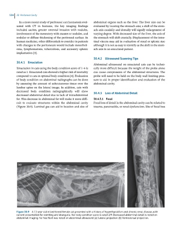
30.4.1 Emaciation
Abdominal ultrasound on emaciated cats can be techni-
Emaciation in cats using the body condition score of 1–6 is cally more difficult because the weight of the probe alone
rated as 1. Emaciated cats showed a higher risk of mortality can cause compression of the abdominal structures. The
compared to cats in optimal body condition [4]. Evaluation probe will need to be held on the body wall limiting pres-
of body condition on abdominal radiographs can be done sure to aid in proper identification and evaluation of the
by assessing the amount of subcutaneous tissue over the abdominal cavity.
lumbar spine on the lateral image. In addition, cats with
decreased body condition radiographically will show
decreased abdominal detail due to lack of intraabdominal 30.4.3 Loss of Abdominal Detail
fat. This decrease in abdominal fat will make it more diffi- 30.4.3.1 Focal
cult to evaluate structures within the abdominal cavity Focal loss of detail in the abdominal cavity can be related to
(Figure 30.9). Luminal gas can aid in location and size of trauma, pancreatitis, or renal dysfunction. Site of focal loss
(b)
(a)
Figure 30.9 A 13-year-old mixed-breed female cat presented with a history of hyperthyroidism and chronic renal disease, with
current presentation for vomiting and stranguria. Her body condition score is rated 2/9. Decreased abdominal detail is noted on
abdominal imaging. No free fluid was noted on abdominal ultrasound. (a) Lateral projection. (b) Ventrodorsal projection.