
Page 521 - Feline diagnostic imaging
P. 521
30.4 Alternate Modalities 533
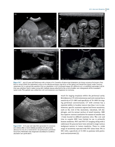
(a) (b)
(c) (d)
Figure 30.7 (a) A 12-year-old Tonkinese with a history of 4–5 months of abdominal distension. (a) A large volume of echogenic free
fluid is noted within the abdominal cavity on the ultrasound image. Aspiration of the spleen and mesenteric lymph nodes revealed
lymphoma. (b) In contrast, the anechoic fluid accumulation in this ultrasound image of a different cat is a modified transudate. (c,d) In
this case, anechoic fluid is noted along with multiple masses attached to the urinary bladder and enlargement of the mesenteric
lymph node. This patient was euthanized and carcinomatosis was diagnosed on necropsy.
much for staging neoplasia within the peritoneal cavity.
Multidetector CT with intravenous and oral contrast has a
sensitivity of 25–100% and specificity of 78–100% for stag-
ing peritoneal carcinomatosis. CT with contrast has a
reported ability to localize tumors less than 1 cm in size.
However, specific anatomic regions had lower sensitivity,
such as the root of the mesentery, omentum, left dia-
phragm, and the serosal surface of small intestine. MRI
has superior contrast resolution for masses of smaller size
<5 mm located in difficult anatomic sites. The cost and
time to acquire MRI have limited its use to primarily
human medicine. PET and PET/CT imaging along with a
radiotracer for glucose have been utilized to stage various
Figure 30.8 A 13-year-old male DLH presented for anorexia malignant tumors in human medicine. There is a wide
and icterus after a recent dental procedure. On abdominal range of sensitivity reported with PET alone from 78% to
ultrasound, free air is noted within the peritoneum just below
the arrows. Peritonitis was diagnosed secondary to duodenal 98% with a specificity of 55–90% in patients with perito-
ulceration and perforation. neal carcinomatosis [2].