
Page 1115 - Cote clinical veterinary advisor dogs and cats 4th
P. 1115
556 Intervertebral Disc Disease
• Neurologic exam (p. 1136)
○ Findings are consistent with a single focal
VetBooks.ir ○ Deficits and segmental reflexes help to
spinal cord lesion (i.e., mentation and
cranial nerve responses are normal)
localize the lesion.
• UMN signs in forelimbs and hindlimbs: 50 mm
C1-C5 lesion
• Lower motor neuron signs in forelimbs,
UMN signs in hindlimbs: C6-T2 (cervical
intumescence) lesion
• Forelimbs normal, UMN signs in hindlimbs: R L
T3-L3 lesion
• Forelimbs normal, decreased patellar reflex:
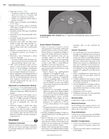
L4-6 lesion INTERVERTEBRAL DISC DISEASE Axial CT image shows intervertebral disc extrusion (arrow) at T12-13
• Forelimbs normal, decreased withdrawal in on the left side.
hindlimbs: L6-S2 lesion
• Cutaneous trunci reflex may help further
localize in dogs with signs referable to a
T3-L3 segment lesion, and might also Acute General Treatment neurologic signs are also candidates for
help determine which side is preferentially • Choice of treatment is based on severity surgery.
affected. and progression of neurologic dysfunction
• Palpation of epaxial muscles and vertebrae and discomfort as well as on owner wishes. Chronic Treatment
to determine presence and location of Discuss referral to a veterinary neurologist or • For animals that have lost motor function,
discomfort surgeon for patients with paresis or paralysis. the single most important aspect of treatment
• Determine a line of decreased or absent • Patients with discomfort alone and no is bladder management to reduce the risk of
sensation, if possible. neurologic deficits or those with mild urinary tract infection and bladder detrusor
○ Location of deficits helps determine the neurologic deficits that are not progressing muscle damage from chronic overdistention.
site of the lesion but does not confirm that rapidly: STRICT cage rest for 4-6 weeks (only This may entail manual expression of the
intervertebral disc disease is the cause. activity is short walks to urinate/defecate) bladder every 4-6 hours or catheterization. In
• CBC, serum biochemistry panel, and uri- regardless of perceived improvement some cases, medication to help decrease ure-
nalysis to assess anesthetic risk; American • Treatment with muscle relaxants and anal- thral sphincter tone (e.g., phenoxybenzamine
Society of Anesthesiologists classification gesics (nonsteroidal antiinflammatory drugs 0.25 mg/kg PO q 12h, avoid if hypotensive;
system (p. 1196). Urinary tract infection [NSAIDs], opiates) prazosin 0.07 mg/kg PO q 8-12h) may be
may accompany dysuria/urine retention. ○ Muscle relaxant (e.g., methocarbamol helpful.
• Survey radiography with orthogonal views of [Robaxin] 15-20 mg/kg PO q 8h • Reduce antiinflammatory medications and
heavily sedated, properly positioned patient ○ NSAIDs: meloxicam 0.1 mg/kg IV, SQ, analgesics after the acute phase, based on
will rule out fractures/luxations and severe or PO q 24h, or carprofen 2.2 mg/kg PO patient comfort.
bone neoplasia. Calcified discs in situ are q 12h or 4.4 mg/kg q 24h, or deracoxib • Confined, padded rest areas; slings for assisted
abnormal (degenerated) but may not be 1-2 mg/kg PO q 24h. Do not use more ambulation; hydrotherapy for cleanliness and
clinically significant. than one NSAID at a time or an NSAID to stimulate ambulation
concurrently with glucocorticoids (risk of • Physical rehabilitation including underwa-
Advanced or Confirmatory Testing gastric ulceration). ter treadmill use to strengthen axial and
• Myelography, CT, CT combined with myelog- • Glucocorticoids appendicular musculature and help retrain
raphy, or MRI (p. 1132) may reveal location of ○ Traditional but increasingly unpopular the nervous system
spinal cord compression. MRI is the preferred with neurosurgeons due to frequent adverse • Acupuncture may be helpful.
method. effects and lack of proven efficacy; higher • Chiropractic maneuvers are controversial and
• Cerebrospinal fluid (CSF) analysis may be success rates reported for NSAID therapy may worsen neurologic status.
done in conjunction with myelography or ○ Methylprednisolone sodium succinate
after CT or MRI, especially if a disorder (Solu-Medrol) 10-30 mg/kg IV once, Nutrition/Diet
other than disc disease is suspected to be within 12 hours of spinal cord injury; Control body weight to avoid obesity.
the cause of myelopathy (pp. 1080 and prednisone 0.5 mg/kg PO q 24h for
1323). 1-3 days; dexamethasone 0.1-0.2 mg/kg Behavior/Exercise
• Advanced imaging is usually done when once Avoid climbing and jumping activities that
surgery is contemplated as a possible ○ Dexamethasone has been associated with extend the spine during and after recovery to
treatment. If a client has ruled out surgical the highest incidence of adverse effects in reduce risk of recurrence.
intervention, the results of any imaging study treatment of IVDD.
are of academic value only and may have little • If acutely paraplegic/tetraplegic: IV fluids to Drug Interactions
influence on further treatment planning or maintain hydration for optimal spinal cord Concurrent use of glucocorticoids and NSAIDs
prognostication. perfusion can lead to severe gastrointestinal ulceration
• Patients with more severe neurologic signs and is contraindicated.
TREATMENT (i.e., non-ambulatory paraparesis, loss of
motor function, and loss of nociception) and Possible Complications
Treatment Overview evidence of cord compression on imaging • Urinary tract infection from incontinence/
Goals of treatment are to alleviate discomfort should have decompressive surgery. urinary retention or improper catheter use/
and pain and reverse neurologic dysfunction • Patients with recurrent signs of neurologic care
by reducing swelling and compression of the dysfunction, uncontrolled spinal pain despite • Detrusor atony from chronic overdistention
spinal cord. strict medical management, and worsening of the bladder
www.ExpertConsult.com