
Page 1120 - Cote clinical veterinary advisor dogs and cats 4th
P. 1120
558 Intracranial Neoplasia
vascular structure responsible for the forma- behavioral change, contralateral hemiparesis, Initial Database
tion of cerebrospinal fluid (CSF) • Brainstem tumors: ipsilateral cranial nerve • CBC, serum biochemistry profile, urinalysis:
altered mental status
VetBooks.ir • Pituitary tumor: tumor of the pituitary deficits, hemiparesis or tetraparesis or • Thoracic and abdominal radiographs: often
• Ependymoma: tumor of the ependymal cells
usually normal
that line the ventricular system
tetraplegia, altered mental status, central
normal but performed to rule out extracranial
gland, called macroadenoma when diameter
vestibular dysfunction
> 1 cm • Cerebellar tumors: hypermetria, intention neoplasia
• Other neoplasms possible: primary CNS tremors, truncal sway, broad-based stance, Advanced or Confirmatory Testing
or generalized lymphoma, histiocytic paradoxical vestibular dysfunction • CT or MRI (p. 1132)
sarcoma, nasal tumor, osteosarcoma and ○ MRI is markedly superior for soft-tissue
other primary bone tumors of the skull, Etiology and Pathophysiology detail (brain, spinal cord) and has higher
metastatic tumors (e.g., hemangiosarcoma, • Cause is unknown. resolution than CT. As a result, this is the
melanoma, mammary tumor and others) • Usually occur as solitary masses, but multiple current gold standard for brain imaging.
tumors can be seen with metastatic disease. ○ CT is an adequate imaging modality,
HISTORY, CHIEF COMPLAINT • Most commonly reported in the rostrotento- is superior for bone lesions (e.g., skull
• Intracranial neoplasia should be considered rial compartment (rostral to the tentorium tumors), is much faster, and is typically
in any older patient with an insidious onset cerebelli, including the cerebrum and less expensive than MRI.
of slowly progressive neurologic signs and in diencephalon) ○ Focal mass identified for most brain
any patient older than 5 years with a recent • Biological behavior (benign vs. malignant) is tumors; however, diffuse neoplasia is
onset of seizures. generally irrelevant because neoplasia of the possible (e.g., lymphoma) and multiple
• Historic findings depend on lesion location. brain is detrimental due to space-occupying tumors may be identified with metastatic
Clinical signs are often insidious and progres- effects. neoplasia.
sive; however, acute onset of clinical signs is ○ CT and MRI features vary between tumor
possible. DIAGNOSIS types.
• The most common chief complaints include • CSF analysis (p. 1080)
seizures, circling, behavior change (aggres- Diagnostic Overview ○ Used as an adjunct to advanced imaging,
sion), altered consciousness, and nonspecific Neoplasia of the brain is suspected based on primarily to rule out encephalitis
signs such as inappetence, lethargy, and signalment, history, and neurologic exam results. ○ With brain neoplasia, results are generally
inappropriate elimination. Confirmation requires advanced intracranial nonspecific and reveal normal to mildly
imaging with CT or MRI. elevated protein.
PHYSICAL EXAM FINDINGS ○ Albuminocytologic dissociation (elevated
• Neurologic exam findings vary, depending Differential Diagnosis CSF protein with normal nucleated cell
on lesion location (p. 1136). • Infectious diseases (bacterial, viral, fungal, count) can occur but is not pathognomonic.
• Findings generally reflect a focal lesion protozoal), depending on geographic location ○ Neoplastic cells are rarely identified in
with asymmetric clinical signs. However, • Inflammatory diseases (e.g., granulomatous the CSF; however, finding lymphoblasts
a large case series found that one-half of meningoencephalomyelitis, necrotizing in the CSF supports a diagnosis of CNS
canine brain tumors occupy more than one encephalitis) lymphoma.
anatomic region of the brain (e.g., forebrain • Cerebrovascular infarction if clinical signs • Histopathologic analysis of tissue is required
and brainstem). are acute, nonprogressive, and asymmetrical for definitive diagnosis. Tissue samples can be
• Cerebral tumors: seizures, contralateral • Toxin and metabolic diseases if clinical signs obtained by surgical excision or stereotactic
menace and postural reaction deficits, are acute, nonprogressive, and symmetrical brain biopsy.
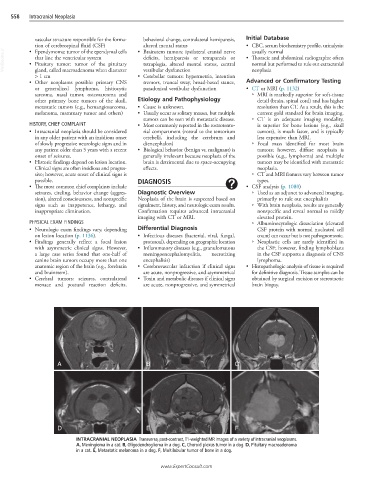
A B C
D E F
INTRACRANIAL NEOPLASIA Transverse, post-contrast, T1-weighted MR images of a variety of intracranial neoplasms.
A, Meningioma in a cat. B, Oligodendroglioma in a dog. C, Choroid plexus tumor in a dog. D, Pituitary macroadenoma
in a cat. E, Metastatic melanoma in a dog. F, Multilobular tumor of bone in a dog.
www.ExpertConsult.com