
Page 1123 - Cote clinical veterinary advisor dogs and cats 4th
P. 1123
560 Intraocular Neoplasia
retrievers, suspected in golden retrievers, • Extension of mass through sclera (indicates in humans, in whom most are of choroidal
more invasive tumor)
origin.
German shepherds • Diffuse hyperpigmentation of iris (more often • Primary intraocular sarcoma in cats occurs
VetBooks.ir ASSOCIATED DISORDERS • Dyscoria, anisocoria months to years after chronic uveitis or
in cats)
Uveitis, glaucoma, retinal detachment, and
blunt or penetrating trauma to the eye.
intraocular hemorrhage can occur secondary
• Hyphema
to intraocular neoplasia. • Shallow anterior chamber Damage to lens epithelium is impli-
cated as initiating factor. Metastasis is
• Displacement of lens (p. 581) common.
Clinical Presentation Findings associated with secondary glaucoma/ • Primary tumors in dogs are benign and,
HISTORY, CHIEF COMPLAINT uveitis: although locally invasive, rarely metastasize.
• Change in appearance of the eye: redness, • Elevated intraocular pressure (glaucoma) • Ciliary body tumors rarely metastasize.
cloudiness, swelling, darkening of iris, mass • Fixed, pupil • Uveal melanoma in cats may metastasize.
noted • Corneal edema • Primary intraocular sarcoma in cats frequently
• Pain, blepharospasm • Scleral injection metastasizes.
• Loss of vision • Hyphema
• Aqueous flare DIAGNOSIS
PHYSICAL EXAM FINDINGS • Lens luxation
Findings directly associated with tumor: • If chronic glaucoma, retinal degeneration, Diagnostic Overview
• Mass that may be pigmented (melanoma/ optic nerve atrophy, and peripapillary (area Goals of diagnosis are to confirm neoplasia
melanocytoma) or pink in color (more around the optic disc) hyperpigmentation versus other processes and differentiate primary
likely ciliary body adenoma/adenocarcinoma from secondary and malignant from benign
or metastatic neoplasia) within the iris or Etiology and Pathophysiology intraocular neoplasm.
pupil • Proliferation of uveal melanocytes or ciliary
• Not all melanomas are pigmented, nor are body epithelium, cause unknown Differential Diagnosis
all pigmented masses melanomas (requires • Melanoma originates most commonly in the • Iris cyst
histologic diagnosis). anterior uvea (iris and ciliary body), unlike • Diffuse iris melanocytosis, hyperpigmentation
1
2
A B
C D E
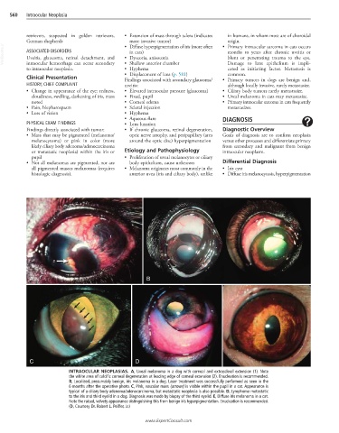
INTRAOCULAR NEOPLASIAS. A, Uveal melanoma in a dog with corneal and extrascleral extension (1). Note
the white area of calcific corneal degeneration at leading edge of corneal extension (2). Enucleation is recommended.
B, Localized, presumably benign, iris melanoma in a dog. Laser treatment was successfully performed as seen in the
6 months after the operative photo. C, Pink, vascular mass (arrows) is visible within the pupil in a cat. Appearance is
typical of a ciliary body adenoma/adenocarcinoma, but metastatic neoplasia is also possible. D, Lymphoma metastatic
to the iris and third eyelid in a dog. Diagnosis was made by biopsy of the third eyelid. E, Diffuse iris melanoma in a cat.
Note the raised, velvety appearance distinguishing this from benign iris hyperpigmentation. Enucleation is recommended.
(D, Courtesy Dr. Robert L. Peiffer, Jr.)
www.ExpertConsult.com