
Page 1126 - Cote clinical veterinary advisor dogs and cats 4th
P. 1126
562 Intussusception
PHYSICAL EXAM FINDINGS Advanced or Confirmatory Testing ○ Resection and anastomosis if unable to
• Palpable, sausage-shaped abdominal mass • Upper GI contrast radiographic study (p. reduce intussusception, a mass is present,
VetBooks.ir palpation of mass may be precluded by for suspected ileocolic intussusception: filling • For enteroenteric intussusception, perform
or there is nonviable bowel
1098), barium enema, or pneumocologram
is characteristic but may not be present;
defect caused by intussusceptum seen within
enteroplication if recurrence appears likely
abdominal guarding/pain.
• Signs of pain on abdominal palpation
intussuscipiens
• Dehydration, tachycardia (more severe signs • Ultrasonography: concentric rings in trans- based on inability to correct underlying
disease.
associated with more proximal obstruction) verse plane (target sign) and hyperechoic • For GEI, perform gastropexy of fundus and
• Poor body condition can be seen in chronic or hypoechoic parallel lines in longitudinal pylorus to prevent recurrence.
cases. views are characteristic. G-shaped or semilu-
• Intussusception may protrude from anus. nar hyperechoic center (mesenteric fat) and Chronic Treatment
visualization of the inner intussusceptum • Opioid analgesia after surgery may decrease
Etiology and Pathophysiology differentiate intussusception from other immediate risk of recurrence.
• Proposed cause is structural or functional conditions. If mesenteric blood flow is • Treat infectious enteritis that may have caused
heterogeneity in the bowel wall, resulting identified with Doppler, the intussusception intussusception.
in an alteration of intestinal pliability or is more likely reducible at surgery. • Antibiotics should be continued after surgery
motility. if peritonitis is present.
• Intussusception produces partial or complete TREATMENT • Enteroplication if not performed at first
intestinal obstruction. surgery and postoperative intussusception
• Increased intraluminal pressure and kinking Treatment Overview recurs
causes collapse of mesenteric blood vessels. Immediate surgical intervention is indicated
Avulsion of vessels can also occur. after hypovolemia is corrected. At surgery, an Possible Complications
• Bowel wall becomes edematous and may attempt is made to reduce the intussusception. • Recurrence of intussusception occurs in up
become ischemic. If it is not reducible or there is bowel damage, to 20% of patients.
• Necrosis of the bowel wall, with leakage of resection and anastomosis are indicated. • Ileus
contents contained by a fibrin seal between Enteroplication may be considered to prevent • Peritonitis associated with bowel rupture
the layers of the intussusception, may occur. If recurrence. Intestinal biopsy is indicated, • Leakage or dehiscence of intestinal suture
leakage is not contained, peritonitis develops. particularly in older patients to evaluate for line
inflammatory bowel disease and neoplasia. • Entrapment and strangulation of bowel
DIAGNOSIS between enteroplication sutures
Acute General Treatment • Foreign body entrapment in bend of intestine
Diagnostic Overview • Intravenous crystalloids to correct dehydration created by enteroplication
The diagnosis of enteroenteric (intestinal) intus- or treat for shock. Colloids may be helpful with • Persistent megaesophagus if GEI (p. 642)
susception is suspected based on history and hypoproteinemia. If severe hypochloremia/
typical physical exam findings. Confirmation hyponatremia, treat with 0.9% NaCl. Potas- Recommended Monitoring
is obtained with abdominal ultrasound exam sium supplementation if hypokalemic • Monitor hydration status and serum elec-
or surgical exploration. GEI is confirmed with • Administer perioperative antibiotics (e.g., trolyte concentrations.
contrast imaging or endoscopy (p. 468). cefazolin 22 mg/kg IV at induction and • Monitor for signs of intestinal suture
q 90 minutes during procedure +/− q 8h line dehiscence and peritonitis (increased
Differential Diagnosis postoperatively) body temperature, abdominal pain,
• Gastroenteritis associated with infection or • Enteroenteric or enterocolic intussuscep- hypoglycemia).
dietary indiscretion tions; at the time of surgical exploration, • If clinical signs recur after surgery, repeat
• Intestinal obstruction associated with foreign reduce intussusception by applying pressure imaging to evaluate for possible recurrence
body, neoplasia, abscess, or granuloma to intussuscipiens while gently pulling on of intussusception or complication associated
• Physiologic ileus intussusceptum. with enteroplication.
• Rectal prolapse if intussusception protruding
through anus (p. 866)
Initial Database
• CBC may show evidence of a stress leuko-
gram or anemia. Increased red blood cell
count may be seen with dehydration.
• Serum chemistry profile may show evidence
of dehydration (increased total protein,
azotemia), hypokalemia, hypochloremia,
hyponatremia, or hypoproteinemia. Alkalosis
may be seen with proximal obstructions.
• Abdominal radiographs may show fluid- or
gas-distended intestinal loops. Tubular mass
effect (sausage shape) of the intussusception
may be seen in the small intestine or in a
gas-filled colon.
• Thoracic radiographs: soft-tissue density within
esophagus if GEI
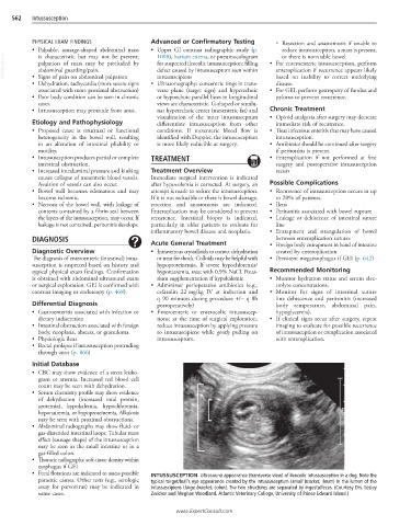
• Fecal flotations are indicated to assess possible INTUSSUSCEPTION Ultrasound appearance (transverse view) of ileocolic intussusception in a dog. Note the
parasitic causes. Other tests (e.g., serologic typical target/bull’s eye appearance created by the intussusceptum (small bracket, ileum) in the lumen of the
assay for parvovirus) may be indicated in intussuscipiens (large bracket, colon). The two structures are separated by ingesta/feces. (Courtesy Drs. Lesley
some cases. Zwicker and Meghan Woodland, Atlantic Veterinary College, University of Prince Edward Island.)
www.ExpertConsult.com