
Page 1687 - Cote clinical veterinary advisor dogs and cats 4th
P. 1687
848 Pustular and Crusting Skin Disorders
Pemphigus is more common in middle-
aged adults.
VetBooks.ir • Drug eruptions can occur at any age.
• Hereditary mechanobullous disease: epider-
molysis bullosa develops shortly after birth.
GENETICS, BREED PREDISPOSITION
• Juvenile cellulitis (p. 567): dachshund,
golden and Labrador retrievers, Gordon
setters, pointers. More than one puppy can
be affected in a litter.
• Superficial suppurative necrolytic dermatitis:
miniature schnauzer
• Pemphigus foliaceus: Akita, chow chow
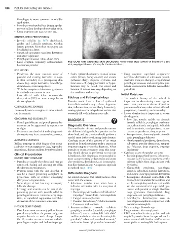
• Drug eruption (especially sulfonamides): PUSTULAR AND CRUSTING SKIN DISORDERS Honey-colored crusts (arrows) on the pinna of a dog
Doberman pinscher with pemphigus foliaceus. (Courtesy Dr. Caroline de Jaham.)
RISK FACTORS
• Pyoderma, the most common cause of • Scales, epidermal collarettes, crusts of various • Drug eruption: superficial suppurative
pustular and crusting dermatitis in dogs, colors (brown, honey colored) and texture necrolytic dermatitis of schnauzers (associ-
is often secondary to a predisposing skin (adherent, flaky), alopecia, erythema, and ated with shampoo therapy), drug-induced
disease (e.g., atopic dermatitis, food allergy, focal areas of hyperpigmentation or hyper- pemphigus foliaceus, and eosinophilic pus-
demodicosis, endocrinopathies). keratosis may be noted. The extent and tulosis (subcorneal to follicular neutrophilic
• With the exception of abscesses, pyoderma location of lesions may vary, depending on pustulosis)
is relatively uncommon in cats. the condition and severity.
• Cats affected with feline immunodefi- Initial Database
ciency virus (FIV) are more susceptible to Etiology and Pathophysiology • The medical history of the animal is
dermatophytosis. Pustules result from a loss of epidermal important in determining cause: age of
intercellular cohesion (e.g., edema, degenera- onset, breed, presence or absence of pruritus,
CONTAGION AND ZOONOSIS tion, inflammation, autoantibody formation), previous medications, other animals affected,
Dermatophytosis is contagious to other animals causing epidermal or subepidermal cavities that progression, chronicity, and seasonality
and is zoonotic. eventually fill with inflammatory cells. • Lesion distribution is important to orient
the diagnosis.
GEOGRAPHY AND SEASONALITY DIAGNOSIS ○ Face (lips, muzzle, eyelids, ear pinnae):
• Pemphigus foliaceus and pemphigus erythe- juvenile cellulitis, pemphigus erythema-
matosus can be aggravated by exposure to Diagnostic Overview tosus, demodicosis, eosinophilic folliculitis
sunlight. The combination of crusts and pustules narrows and furunculosis, nasal pyoderma, muco-
• Pyodermas associated with underlying atopic the differential diagnosis, but pustules can be cutaneous pyoderma, drug eruption
dermatitis may have a seasonal occurrence. short-lived, and the clinician should perform a ○ Feet: pyoderma, dermatophytosis, demodi-
careful exam before confirming their absence. cosis, pemphigus foliaceus
ASSOCIATED DISORDERS Cytologic exam of the content of an intact ○ Trunk: superficial pyoderma, demodicosis,
Bullous impetigo in adult dogs is often associ- pustule or from the exudates under a crust is an subcorneal pustular dermatosis, pemphi-
ated with immunosuppression (e.g., hyperadre- important step to orient the diagnosis. When gus foliaceus, drug eruption, impetigo
nocorticism, diabetes mellitus, hypothyroidism). pustules or crusts are seen on dogs, skin scrap- (abdomen)
ings should always be performed to rule out • Cytologic exam of pustular contents
Clinical Presentation demodicosis. Skin biopsies are recommended in ○ Bacteria: phagocytized bacteria (often cocci
HISTORY, CHIEF COMPLAINT most cases presenting with pustules and crusts because staphylococcal organisms are the
• Pustules are usually short-lived and may go after pyoderma, demodicosis, and dermatophy- primary isolates from dogs and cats with
unnoticed. Scaling and crusting are often tosis have been ruled out. A diagnostic approach pyoderma)
the dominant presentation. is outlined on p. 1446. ○ Neutrophils: pyoderma, pemphigus
• Pruritus varies with the skin disorder. It complex, subcorneal pustular dermatosis,
can be a major presenting complaint in Differential Diagnosis and canine linear IgA pustular dermatosis
pyodermas, with or without underlying An asterisk indicates that intact pustules often ○ Eosinophils: abundant eosinophils with
allergies or ectoparasitism. occur with the disease. eosinophilic folliculitis/furunculosis and
• Some degree of hair loss may accompany • Infectious: pustules most often have a sterile eosinophilic pustulosis; eosinophils
follicular damage. follicular orientation with the exception of are also associated with superficial pyo-
• Lethargy and anorexia can be part of the impetigo. dermas with parasitic or allergic disorders,
presenting picture with juvenile cellulitis, ○ Bacterial: pyoderma (bacterial folliculitis,* deep pyodermas (furunculosis), drug
pemphigus foliaceus, cutaneous drug erup- impetigo,* furunculosis), dermatophilosis eruptions, and pemphigus foliaceus.
tions, or superficial suppurative necrolytic ○ Fungal: dermatophytosis ○ Acantholytic keratinocytes: seen in
dermatitis of the miniature schnauzer. ○ Parasitic: demodicosis,* Pelodera dermatitis pemphigus complex in conjunction with
○ Protozoal: leishmaniasis numerous neutrophils
PHYSICAL EXAM FINDINGS • Immune-mediated: juvenile cellulitis, • Skin scrapings: Demodex spp
• Pustules are most commonly yellow. Green pemphigus complex (especially pemphigus • Fungal culture: dermatophytosis
pustules may indicate the presence of gram- foliaceus*), canine eosinophilic folliculitis* • CBC, serum biochemistry profile, and uri-
negative bacteria or toxic change. Larger, and furunculosis, canine sterile eosinophilic nalysis if systemic disease is suspected; results
flaccid pustules are more common with the pustulosis,* subcorneal pustular dermatosis,* often unremarkable besides mild/moderate
pemphigus complex and bullous impetigo. and canine linear IgA pustular dermatosis* neutrophilic or eosinophilic leukocytosis
www.ExpertConsult.com