
Page 1965 - Cote clinical veterinary advisor dogs and cats 4th
P. 1965
Tooth Fractures 981
• Root fracture: fracture involving the DIAGNOSIS invite infection of endodontic and periapical
root Diagnostic Overview tissues.
VetBooks.ir HISTORY, CHIEF COMPLAINT A sedated/anesthetized oral exam and dental Acute General Treatment Diseases and Disorders
• Uncomplicated crown fractures may not
radiography are needed to determine whether
• Tooth fractures commonly noted as incidental
findings on routine physical exam
the rough tooth surface and sealing dentinal
root.
• History of falling from a height, vehicular the fracture is complicated or involves the require treatment or benefit from smoothing
trauma, fights with other animals, aggressive tubules of exposed dentin with a bonding
chewing tendencies, or other trauma Differential Diagnosis agent.
• Abrasion/attrition • Acute complicated crown fracture: vital pulp
PHYSICAL EXAM FINDINGS • Tooth resorption therapy (partial pulpectomy, direct pulp
• Commonly no overt clinical signs, especially • Caries capping, and restoration) within 48 hours
if fracture is uncomplicated • Displacement injury (luxation/avulsion) of pulp exposure. Administer antibiotics
• Oral bleeding with complicated fractures (e.g., intraoperative ampicillin 22 mg/kg IV
• Hypersalivation with acute complicated Initial Database q 6h, followed by amoxicillin/clavulanic acid,
fractures CBC, serum chemistry panel, urinalysis: gener- 14 mg/kg PO q 12h × 7 days, or clindamycin
• Appetite rarely affected; chewing on opposite ally unremarkable (preanesthetic) 11 mg/kg PO q 12h × 7 days).
side resulting in greater calculus accumula- • Extraction of fractured deciduous or perma-
tion on affected side Advanced or Confirmatory Testing nent teeth
• Calculus may obscure a slab fracture of the • Anesthetized oral exam using a dental
maxillary fourth premolar; compare with explorer (#11/12, 17, or 23) to determine Chronic Treatment
crown height and shape of the contralateral pulp exposure (p. 1140) • Chronic, complicated crown fractures (adult
tooth. • Dental radiographs are indicated to identify animals): root canal therapy or extraction
• Acute pulp exposure: red spot in tooth defect extent of lesion and state of adjoining teeth; • Fractures extending below the gingival
± bleeding from exposed pulp, painful on possible findings include structural crown attachment (crown-root fractures) may
probing and/or root defect, arrested root development require periodontal surgery to prevent focal
• Chronic pulp exposure: dark brown/black (open root apex, wide root canal compared periodontal pocketing.
spot in tooth defect, asymmetrical calculus with healthy, contralateral tooth), diffuse root • Prosthodontic crowns are indicated after root
accumulation (greater on affected side), canal mineralization, apical root resorption, canal therapy to protect the remainder of
regional facial swelling ± draining tracts at and/or periapical lucency. the crowns in dogs.
mucogingival junction or through skin
TREATMENT Possible Complications
Etiology and Pathophysiology • Failure of endodontic therapy: repeat root
• Uncomplicated fracture: exposure of dentinal Treatment Overview canal therapy, perform apicoectomy and
tubules causes sensitivity and may allow Endodontic therapy or extraction is indicated retrograde filling, or extract the tooth.
bacterial access to the pulp. Odontoblasts for fractured teeth with pulp exposure. Frac- • Uncontrolled force during tooth extraction
may respond by forming tertiary dentin, tured teeth with no pulp exposure require ○ Fracture and incomplete removal of the
sealing off exposed tubules. radiographic evaluation and monitoring because tooth/root, resulting in periapical abscess
• Complicated fracture: pulp exposure results they may also develop endodontic disease. A (p. 7)
in pulpitis and bacterial infection; most wait-and-see approach for fractured teeth with ○ Trauma to soft tissues (eye, brain, tongue,
develop pulp necrosis and periapical disease pulp exposure is below the standard of care salivary gland ducts, and neurovascular
(p. 7). because tooth fractures cause discomfort and structures)
*
A B
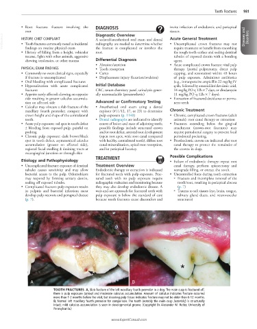
TOOTH FRACTURES A, Slab fracture of the left maxillary fourth premolar in a dog. The main cusp is fractured off;
there is pulp exposure (arrow) and moderate calculus accumulation. Amount of calculus indicates fracture occurred
more than 1-2 months before the visit, but bleeding pulp tissue indicates fracture may not be older than 6-12 months.
B, Normal left maxillary fourth premolar for comparison. The tooth (notably the main cusp [asterisk]) is structurally
intact; mild calculus accumulation is seen in developmental groove. (Copyright Dr. Alexander M. Reiter, University of
Pennsylvania.)
www.ExpertConsult.com