
Page 2013 - Cote clinical veterinary advisor dogs and cats 4th
P. 2013
1007.e2 Urachal Diverticulum
Urachal Diverticulum
VetBooks.ir
○ May be associated with or predispose the
BASIC INFORMATION
opaque uroliths
animal to chronic/recurrent UTI and • Abdominal radiographs to rule out radi-
Definition urolithiasis
Embryonic remnant at the apex of the urinary • Microscopic diverticulum Advanced or Confirmatory Testing
bladder; although diverticula occur commonly, ○ Most common form in cats; can be • Contrast cystography
they seldom result in clinical problems identified in up to 40% ○ Positive-contrast cystography
○ Remnants of urachus at bladder apex ○ Double-contrast cystography
Synonym that can extend from the level of the • Ultrasonography
Vesicourachal diverticulum submucosa to the subserosa • Cystoscopy
○ Not associated with clinical signs • Exploratory celiotomy and cystotomy
Epidemiology • Acquired macroscopic diverticulum
SPECIES, AGE, SEX ○ Microscopic diverticula may become TREATMENT
Dogs and cats, both sexes macroscopic secondary to sustained
increase in bladder intraluminal pressure. Treatment Overview
RISK FACTORS ○ May spontaneously regress if the Incidentally discovered urachal diverticula typi-
Microscopic diverticula in cats may create risk cause of increased bladder pressure is cally do not require therapy. Those associated
for developing macroscopic diverticula after removed with recurrent UTI or urolithiasis are surgically
urinary tract obstruction from any cause. addressed.
DIAGNOSIS
ASSOCIATED DISORDERS Acute General Treatment
• Urinary tract infection (UTI)/bacterial cystitis Diagnostic Overview • Relieve urethral obstruction if present.
• Urolithiasis (especially struvite) Diagnosis of urachal diverticulum is based on • Fluid therapy and correction of electrolyte
• Feline lower urinary tract signs/disease imaging of the urinary tract through ultrasonog- disturbances if present
(FLUTS/D) raphy, contrast urethrocystography, cystoscopy, • Antimicrobial therapy for bacterial cystitis
or visual inspection at surgery. Often, diverticula if present
Clinical Presentation are incidental findings.
DISEASE FORMS/SUBTYPES Chronic Treatment
• Macroscopic: intramural and extramural Differential Diagnosis • Animals with clinical signs or a UTI related
• Microscopic • Neoplasia to congenital macroscopic diverticula or non-
• Acquired macroscopic • Polyps resolving acquired macroscopic diverticula
• Urolithiasis should undergo surgical resection of the
HISTORY, CHIEF COMPLAINT • Blood clots diverticulum:
Clinical signs are often absent. When clinical ○ Exploratory celiotomy
signs are apparent, they may include any of Initial Database ○ Ventral midline cystotomy
the following: • CBC: unremarkable ○ Identification of diverticulum at apex of
• Hematuria • Serum chemistry profile: unremarkable unless bladder
• Pollakiuria urethral obstruction exists ○ Excision of diverticulum with elliptical
• Dysuria • Urinalysis: often unremarkable; sometimes incision
• Stranguria shows pyuria, hematuria, bacteruria, or ○ Routine closure
• Inappropriate elimination struvite crystalluria • Address chronic feline lower urinary tract
• Systemic illness due to urinary tract obstruc- • Urine culture and sensitivity (C&S) testing signs/disease.
tion (rare) is used for identifying UTI. • Address urolithiasis.
PHYSICAL EXAM FINDINGS
Physical exam is usually unremarkable. When
present, abnormalities are nonspecific:
• Hematuria (stains on prepuce, vulva, or
hocks)
• Painful urinary bladder
• Enlarged, turgid bladder with urethral
obstruction
Etiology and Pathophysiology
The urachus is a canal connecting the fetal
bladder with the allantois. The urachal lumen
normally becomes obliterated during develop-
ment, but on occasion the lumen remains patent
(patent urachus) or obliteration is incomplete,
leaving a remnant diverticulum. Cause of
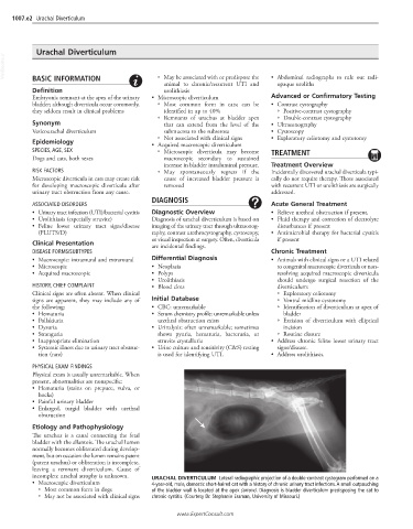
incomplete urachal atrophy is unknown. URACHAL DIVERTICULUM Lateral radiographic projection of a double-contrast cystogram performed on a
• Macroscopic diverticulum 4-year-old, male, domestic short-haired cat with a history of chronic urinary tract infections. A small outpouching
○ Most common form in dogs of the bladder wall is located at the apex (arrow). Diagnosis is bladder diverticulum predisposing the cat to
○ May not be associated with clinical signs chronic cystitis. (Courtesy Dr. Stephanie Essman, University of Missouri.)
www.ExpertConsult.com