
Page 2312 - Cote clinical veterinary advisor dogs and cats 4th
P. 2312
1150 Pericardiocentesis
○ Do not discontinue PN abruptly, espe- Alternatives and Their
cially in patients that are not eating. If a Relative Merits
VetBooks.ir • Catheter care • In general, nutrient delivery by the enteral
patient cannot be weaned, monitor for
Enteral nutrition:
hypoglycemia.
route is safer and less costly than PN.
○ Examine the catheter site at least daily for
tract and supports the structure and function
signs of phlebitis or infection, changing • Enteral nutrition has trophic effects on the GI
the dressing as needed. of the mucosa and the barrier function of
○ Change the drip set every day. the gut.
○ Never use the catheter for any purpose • Preferred over PN whenever feasible
other than the delivery of PN (e.g., no
blood sampling, administering medica- SUGGESTED READING
tions or fluids). Michel KE, et al: Parenteral nutrition. In Silverstein
• Monitoring DC, et al, editors: Small animal critical care medi-
○ In addition to the routine monitoring cine. ed 2, St. Louis, 2015, Elsevier, pp 687-690.
appropriate for any animal on IV fluid AUTHOR: Kathryn E. Michel, DVM, MS, MSED,
therapy, patients receiving PN should be DACVN
monitored for metabolic complications. EDITORS: Leah A. Cohn, DVM, PhD, DACVIM; Mark S.
○ Blood glucose monitored at least q 12h Thompson, DVM, DABVP
and serum electrolytes at least q 24h
○ When a packed cell volume (PCV) test is
performed, the serum should be examined
for evidence of lipemia.
○ CBC and serum biochemistry profile as
indicated and at least once weekly
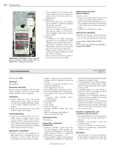
PARENTERAL NUTRITION Infusion pump and
prepared parenteral nutrition solution ready for
administration. (Courtesy Dr. Lesley King.)
Pericardiocentesis Bonus Material
Online
Difficulty level: ♦♦ • Clippers, surgical scrub, isopropyl alcohol, benefit of pericardiocentesis in relieving cardiac
and gauze pads for clipping and preparing tamponade outweighs the risks.
Synonym the skin • For stable patients, cursory echocardiogram
Pericardial tap • Lidocaine 0.5-3 mL for local block +/− for is performed before centesis because cardiac
ventricular arrhythmias tumors are more easily visible with an outline
Overview and Goal • 3-mL syringe for local block of anechoic PE.
Removal of pericardial effusion (PE) for diag- • Ultrasound machine for location selection • A peripheral intravenous (IV) catheter is
nostic investigation and for therapeutic relief • Electrocardiographic (ECG) monitoring placed for sedation, antiarrhythmic doses
1
of signs related to cardiac tamponade • 14-18 gauge, 2- 5 4 inch over-the-needle of lidocaine, or emergency use.
catheter • Continuous ECG monitoring should be
Indications ○ 21-gauge, 1-inch butterfly catheter employed whenever possible, both during
• Cardiac tamponade: when PE elevates preferred in cats and just after pericardiocentesis.
intrapericardial pressure above pressure in • #11 scalpel blade • Assistants are necessary to provide manual
the right heart, resulting in lethargy, collapse • Sterile gloves restraint and should be encouraged to stay
or near collapse, abdominal distention, or • 6-mL syringe vigilant because many dogs feel relief within
dyspnea • 20-35 mL syringe(s) only a few moments of removing PE and
• Diagnostic investigation of the cause of PE • Optional extension tubing and 3-way may attempt to move.
by fluid analysis and cytology stopcock
• Bowl for collecting drained effusate Possible Complications and
Contraindications • Red- and lavender-top tubes Common Errors to Avoid
• Small-volume effusion where the risk of • Ventricular arrhythmias warranting vigilant
cardiac puncture is high (caution in cats Anticipated Time ECG monitoring and ready availability of
because volume of effusion often is small) 10-60 minutes lidocaine.
• Caution in cases of suspected left atrial tears • Re-effusion: risk depends on cause with
(which seldom result in tamponade and may, Preparation: Important variable time frame
with pericardiocentesis, promote continued Checkpoints • Rare risk of chamber or tumor puncture
hemorrhage from the left atrium) • Diagnostic evaluation of the cause of PE causing acute hemorrhage, cardiovascular
(by echocardiogram, fluid analysis and collapse, or death
Equipment, Anesthesia cytology, chest radiographs, CBC, chemistry
• Manual restraint is often sufficient, but profile, urinalysis, T 4 [cats], +/− abdominal Procedure
light sedation (e.g.. butorphanol) may be ultrasound) is encouraged (p. 773). • Restrain patient in left lateral recumbency
warranted to reduce patient movement. • Clients should be educated about potential because catheter entry from the right side
Avoid agents that cause excessive panting. complications but should also be told that the of the thorax is less likely to lacerate lung
www.ExpertConsult.com