
Page 498 - Cote clinical veterinary advisor dogs and cats 4th
P. 498
224 Cryptococcosis
PHYSICAL EXAM FINDINGS often reveals organisms, but complications • Fungal culture and/or histopathology may be
Cats: may be seen after CSF tap due to increased needed to confirm diagnosis in some cases.
VetBooks.ir Swelling or ulcerated mass over the bridge • Anterior uveitis and granulomatous chorio- • Cross-sectional imaging is not usually neces-
intracranial pressure.
Fungal sensitivity testing can be performed
• Upper respiratory tract signs predominate.
when the organism is cultured.
of the nose is common. Stertor or stridor
retinitis should increase suspicion for fungal
may occur with pharyngeal/nasopharyngeal
involvement. Dyspnea from pleural or infection. sary but may increase suspicion of fungal
infection when multifocal mass lesions in
lower airway involvement is less common. Differential Diagnosis the CNS or evidence of meningoencepha-
Submandibular lymphadenopathy is often • Nasal discharge (p. 1255); nasal deformity litis is noted. MRI may reveal meningeal
seen. makes neoplasia the primary differential enhancement along with single or multifocal
• Fundic exam often reveals granulomatous diagnosis contrast-enhancing mass lesions that tend to
chorioretinitis, retinal hemorrhage, retinal • CNS be hyperintense on T2-weighted images and
detachment, and/or evidence of optic ○ Infectious: many, including feline infec- hypointense on T1-weighted images. In cats
neuritis. tious peritonitis, toxoplasmosis, rabies, and dogs with sinonasal disease, nasal mass
• Cutaneous or subcutaneous masses/nodules canine distemper, rabies, ehrlichiosis, lesions or fluid opacification are common
or ulcerated lesions may be noted. Ulcers on Rocky Mountain spotted fever on CT, with contrast-enhancing mass lesions
footpads may result in lameness. ○ Noninfectious: granulomatous meningo- often associated with lysis of the nasal septum
• CNS signs are usually subtle in cats. encephalitis, neoplasia and cribriform plate.
Dogs: • Cutaneous: abscesses, autoimmune skin
• Systemic involvement with findings that disease, neoplastic disease TREATMENT
depend on significance of organ system • Other systemic fungal disease (dogs)
involvement. Treatment Overview
• Ocular, CNS, and cutaneous signs are Initial Database Long-term treatment which azole antifungal
common. • Ocular exam may reveal anterior uveitis and drugs such as fluconazole or itraconazole is
• Fundic exam as in cats. posterior segment changes (granulomatous effective in many infected patients. Surgical
• Severe lethargy and anorexia or obtunded chorioretinitis, retinal detachment, optic neu- cytoreduction may improve treatment success
state may be noted. ritis, retinal hemorrhage, and exophthalmos). if masses can be easily removed.
• Upper respiratory tract signs less common • CBC, serum chemistry panel, and urinalysis:
in dogs. usually minimal change. Normocytic, normo- Acute General Treatment
• Cutaneous and subcutaneous masses and chromic nonregenerative anemia is common. • Although the most effective therapy is
ulcerated lesions may be noted. Mild leukocytosis with neutrophilia and likely amphotericin B in combination with
monocytosis is often seen with eosinophilia 5-flucytosine (cats) or fluconazole (dogs),
Etiology and Pathophysiology likely in cats and a left shift occasionally most patients can initially be treated with
• Caused by a dimorphic fungus of the genus seen in dogs. Mild serum chemistry panel azole monotherapy or a combination of an
Cryptococcus changes reflect organ system involvement. azole and terbinafine.
• Filamentous form in the environment pro- Occasionally, Cryptococcus organisms are • Itraconazole 5-10 mg/kg PO q 24h or flu-
duces basidiospores that are inhaled and form found in urine sediment. conazole 10 mg/kg PO q 12h are the azoles
encapsulated yeast in nasal cavity or lung • Thoracic radiographs are usually normal; of first choice. Fluconazole penetrates the
tissue before disseminating by hematogenous sternal or hilar lymphadenopathy or focal blood-brain and blood-ocular barriers better
route. increases in pulmonary parenchymal opacity than itraconazole and is excreted through the
• C. neoformans and C. gattii cause the majority may be noted in dogs. Rare pleural effusion urinary tract, but studies in humans indicate
of infections in cats, dogs, and humans, but • Abdominal ultrasound is usually normal equal efficacy in CNS infection.
at least 19 species have been implicated. in cats but may reveal mild ascites, lymph-
• Pigeons are considered an important vector; adenopathy, focal/multifocal thickening of
organisms can survive up to 2 years in pigeon the stomach and intestines, and/or masses
roosts and droppings. associated with spleen, kidney, or pancreas
in dogs.
DIAGNOSIS
Advanced or Confirmatory Testing
Diagnostic Overview • Cytology of aspirates of nodules, smears of
• In patients with systemic signs, CBC, nasal discharge, body fluids, or impression
serum chemistry panel, urinalysis, and smears from ulcerated skin lesions may reveal
diagnostic imaging (thoracic radiographs, cryptococcal organisms in 60%-80% of
abdominal ultrasound) will usually be the first cases. Diff-Quik, Romanowsky-type, and
steps. Gram stains can be used to demonstrate
• If nasal involvement is obvious or cutane- round yeast organisms with a large muco-
ous nodules or ulcers are apparent, samples polysaccharide capsule. Granulomatous or
(swabs, fine-needle aspirates, impression pyogranulomatous inflammation is typical.
smears, biopsy) should be collected for • Serologic testing has the advantages of
cytology, culture, and/or histopathology. being inexpensive, highly sensitive and
• Serologic testing for cryptococcal antigen is specific, and noninvasive. Latex agglutina-
an inexpensive and a sensitive and specific tion assays detect capsular antigen and can
test that should be performed early in be used on serum or CSF. Serum antigen
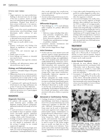
the workup if cytology has not revealed titers are usually very high, but even low CRYPTOCOCCOSIS Cytology shows capsulate
yeasts with narrow-neck budding and prominent
cryptococcal organisms. titers may indicate active infection. Titers unstained region surrounding each yeast (cor-
• CNS signs should result in neurologic exam can be used to follow treatment because they responding to capsule) from the Diff-Quik–stained
with cross-sectional imaging based on neu- decrease significantly in successfully treated smear. (Courtesy Professor Richard Malik, University
rolocalization. Cerebrospinal fluid (CSF) tap animals. of Sydney, Sydney, Australia.)
www.ExpertConsult.com